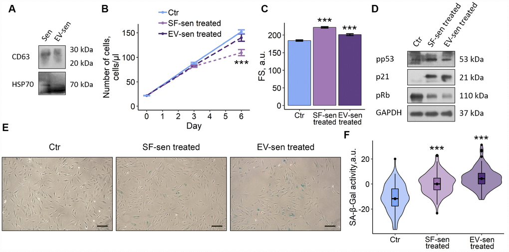
Figure 4.Soluble factors and extracellular vesicles secreted by senescent ESCs trigger senescence in young cells. Sen – senescent ESCs. Ctr – young ESCs cultured in standard conditions. SF-sen or EV-sen treated – young ESCs exposed to soluble factors and extracellular vesicles secreted by senescent ESCs, respectively. (A) Western blot analysis of CD63 and HSP70 total proteins amount in Sen and EV-sen lysates. (B) and (C) Growth curves and cell size of Ctr, SF-sen and EV-sen treated ESCs determined by FACS. Forward scatter (FS) reflects the average cell size evaluated after 6 d of exposure. Values are M ± S.D. (N=3). *** – p<0.005 by ANOVA with Tukey HSD versus Ctr. (D) Western blot analysis of p53 and Rb phosphorylation levels and p21 protein expression performed after 7 d of treatment. Representative results of the three experiments are shown in the Figure. GAPDH was used as loading control. (E) SA-β-Gal staining of Ctr, SF-sen and EV-sen treated ESCs. After 7 d of treatment ESCs were reseeded and additionally cultured for 3 d in order to perform staining of non-confluent cultures. (F) Quantification of SA-β-Gal activity values (E). Values presented as M and 95 % CI (N=100). *** – p<0.005 by ANOVA with Tukey HSD versus Ctr.