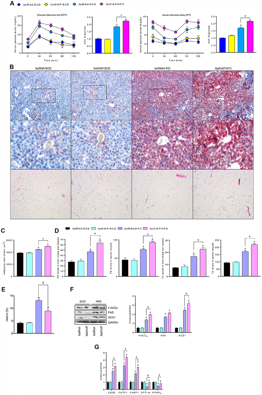
Figure 3.XIAP knockdown aggravates HFD-induced hepatic dysfunction. (A) Glucose tolerance test (GTT) with area under curve (AUC) (Left), and insulin tolerance tests (ITT) with area under curve (AUC) (Right) in mice fed with SCD or HFD were measured. (B) Representative Oil-Red-O-stained images of liver sections (top and medium) and HE-stained adipose tissue (bottom) from each group of mice after SCD or HFD treatment at 12 weeks. (C) The quantification of adipocyte area. (D) Serum lipid (including TG and TC), liver lipid (including TG and TC) levels and glycosylated hemoglobin (E) were assessed. (F) Representative western blot analysis for the expression of p-ACCα, FAS and SCD1 in the livers from each group of mice. (G) qPCR analysis for genes involved in fatty acid metabolism (CD36, FATP1, FABP1, CPT-1α and PPARα) in the livers of mice were performed. For all bar plots shown, data are expressed as the mean ± SEM. n = 8 per group. ap < 0.05 vs. ItsiRNA/SCD or ItsiXIAP/SCD. bp < 0.05 vs. ItsiRNA/HFD.