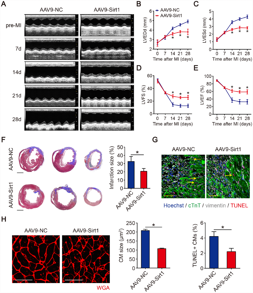
Figure 6.Sirt1 induces cardiac regeneration after MI in adult mice. (A–E) M-mode ultrasonic cardiography changes in mouse hearts, as detected by echocardiography in AAV9-NC and AAV9-Sirt1 adult mice pre-MI and 7, 14, 21, and 28 days after MI. Quantitative analyses were performed for LVEDd (B), LVESd (C), LVFS (D), and LVEF (E) (n=5). (F) Masson’s trichrome staining of serial ventricular sections in AAV9-NC and AAV9-Sirt1 adult mice 28 days after MI. Infarction size was quantified (n=5). Scale bar, 1mm. (G) Cell apoptosis was detected by TUNEL staining and TUNEL-positive CMs were quantified in AAV9-NC and AAV9-Sirt1 adult mice 28 days after MI. Scale bar, 20μm. Quantitative analyses are representative of fields from 5 mice per group. (H) WGA staining and quantitative analyses of CMs size in AAV9-NC and AAV9-Sirt1 adult mice 28 days after MI. Scale bar, 50 μm. Quantitative analyses are representative of fields from 5 mice per group. Statistical significance was calculated using a one-way ANOVA followed by LSD post hoc test in B-E and a two-tailed unpaired Student’s t-test in F-H. *p<0.05; data are presented as the mean ± S.E.M.