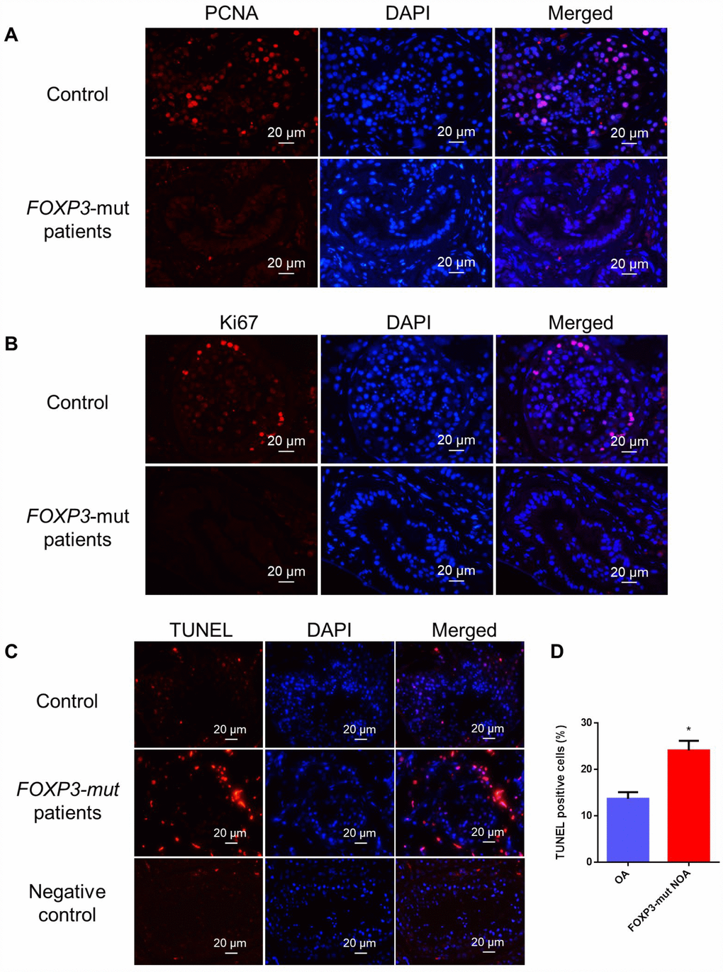
Figure 3.Cell proliferation and apoptosis in the testis of FOXP3-mut NOA patients and OA controls. (A, B) Immunohistochemical staining showed the levels of PCNA (A) and Ki67 (B), the hallmarks for cell proliferation, were decreased in FOXP3-mut NOA patients (lower panels) compared to the OA controls (upper panels). (C) TUNEL assay demonstrated the TUNEL-positive cells (red fluorescence) in FOXP3-mut NOA patients and OA controls. DAPI (blue fluorescence) was used to label cellular nuclei. Replacing the TdT enzyme with PBS was used as the negative control. Scale bars in A-C= 20 μm. (D) The percentages of apoptosis in male germ cells of FOXP3-mut NOA patients and OA controls were calculated using Student’s t-test. All values are means ± SD from three independent experiments. * indicated statistically significant differences (p<0.05).