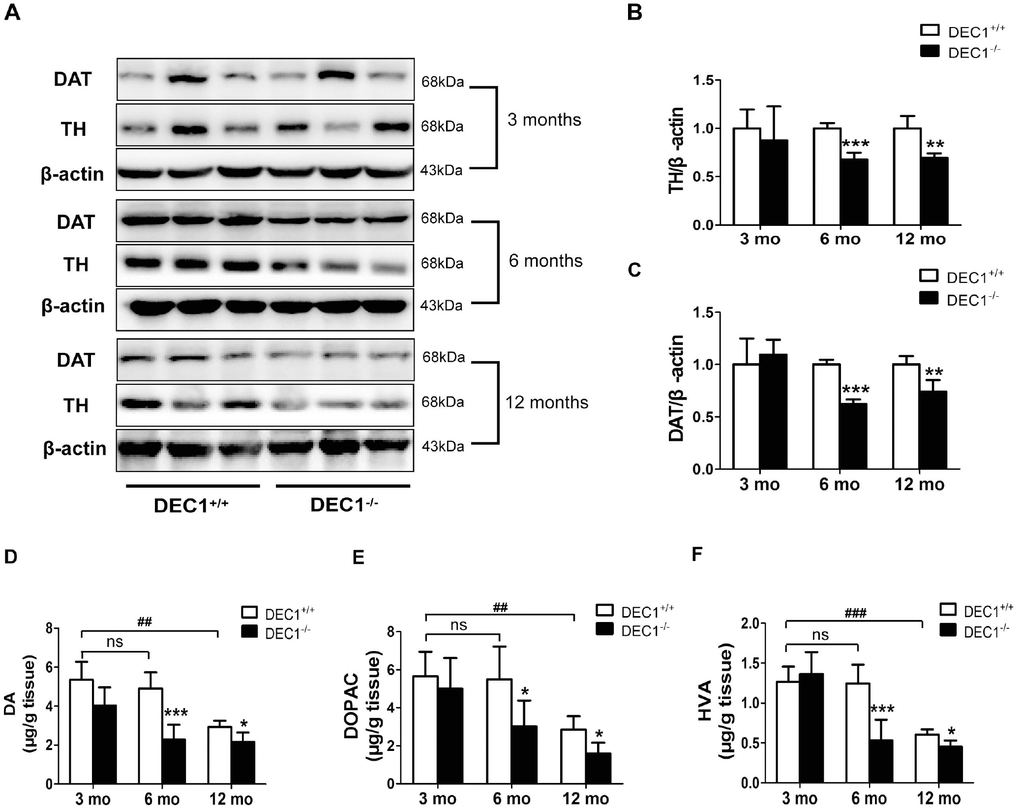
Figure 3.DEC1 deficient mice exhibit a decrease of TH and DA neurons in the midbrain. (A) TH and DAT expression in the midbrain of DEC1+/+ and DEC1-/- mice (n=6 in each group) at the age of 3, 6, 12 months using Western blotting. (B) TH/β-actin (Two-way AONVA, gene: F(1,16)=8.618, p=0.001; age: F(2,16)=0.963, p=0.403 interaction: F(2,16)=3.019, p= 0.077). (C) DAT/β-actin (Two-way AONVA, gene: F(1,18)=9.519, p=0.002; age: F(2,18)=12.897, p=0.02; interaction: F(2,18)=2.832, p= 0.185). (D–F) The amount of dopamine (DA), dihydroxyphenylacetic acid (DOPAC), and homovanillic acid (HVA) in the striatum of DEC1+/+ and DEC1-/- mice at the age of 3, 6, 12 months using HPLC (n=6 in each group). (D) DA (Two-way AONVA, gene: F(1,25)=32.562, p<0.001; age: F(2,25)=16.683, p<0.001; interaction: F(2,25)=4.247, p= 0.272). (E) DOPAC (Two-way AONVA, gene: F(1,25)=9.216, p=0.006; age: F(2,25)=0.963, p<0.001; interaction: F(2,25)=1.373, p= 0.026). (F) HVA (Two-way AONVA, gene: F(1,25)=11.539, p=0.006; age: F(2,25)=33.317, p<0.001; interaction: F(2,25)=10.872, p<0.001). The data are analyzed using t-test for the same age in two genotypes of mice and expressed as mean ± SD. *p<0.05, **p<0.01, ***p<0.001 DEC1-/- mice vs the age-matched DEC1+/+ mice; ##p<0.01, ###p<0.001, ns p>0.05, comparisons are shown in the figure.