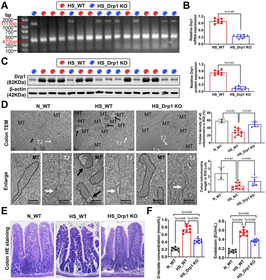
Figure 2.The effects of Drp1 on intestinal barrier function after shock. (A) The genotype identification results of Drp1 KO and WT mice. The genotype of Drp1 KO mice (blue dot, n=8) is Drp1 +/- (double bands at 1115 bp and 472 bp), and the genotype of WT mice (red dot, n=8) is Drp1 +/+ (single band at 472 bp). (B) Drp1 mRNA expression in colon tissues of HS_WT group and HS_Drp1 KO group detected by QRT-PCR (n=8 mice/ group). (C) Drp1 protein expression in colon tissues of HS_WT group and HS_Drp1 KO group detected by Western Blotting (n=8 mice/ group). (D) The mitochondrial morphology and intestinal epithelial tight junctions observed by electronic microscopy (TEM). MT, mitochondria (black arrows refer to vacuolated cristae structure); TJ, tight junctions (white arrows). The boundaries of mitochondria are depicted and the lengths as well as cristae density of all mitochondria in ROI (region of interest) are calculated by Image J software. The mitochondrial number in ROI: 2 in N_WT group, 10 in HS_WT group and 6 in HS_Drp1 KO group. (Bar, 500nm). (E) The intestinal villus morphology observed by immunohistochemistry (400X). (F) The plasma D-lactate and endotoxin concentration in each group (n=8 mice/ group). N_WT, WT mice in normal condition; HS_WT, WT mice in hemorrhagic shock condition; HS_Drp1 KO, Drp1 KO mice in hemorrhagic shock condition. a represents p < 0.05 compared with N_WT group; b represents p < 0.05 compared with HS_WT group.