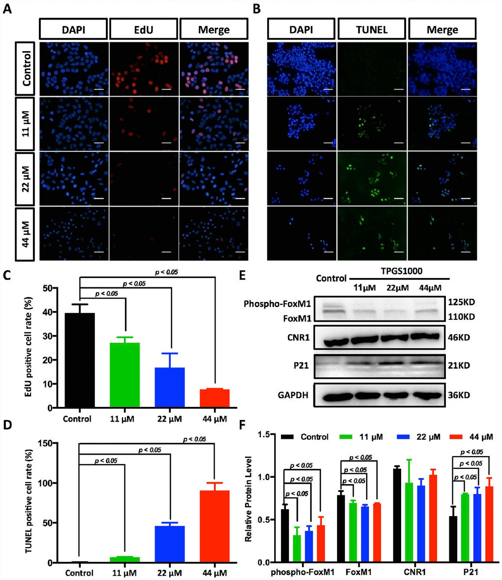
Figure 4.Suppression of DNA synthesis and induction of apoptosis in TPGS-treated HCC cells. (A) Detection by fluorescence microscopy of EdU (red) incorporated into the DNA of cultured HCC cells, scale bar = 40 μm. The nuclei were counter-stained with DAPI (blue). (B) TUNEL (green) positive apoptotic cells in HCC cells induced by TPGS treatments, scale bar = 20 μm. (C) The rates of EdU positive cells that passed through the S phase were calculated with ImageJ, and the 44 μM TPGS group had the lowest EdU positive cell rate (7%). (D) The rates of TUNEL positive cells were elevated with increasing TPGS concentrations, and the 44 μM TPGS group had the highest apoptotic cell rate (approximately 93%). (E) A decrease of FoxM1 and phosphorylated FoxM1, and an increase of p21 protein levels in TPGS-treated HCC cells. (F) Quantitative analysis of western blot results from (E). All protein levels were normalized with the housekeeping genes GAPDH and β-actin.