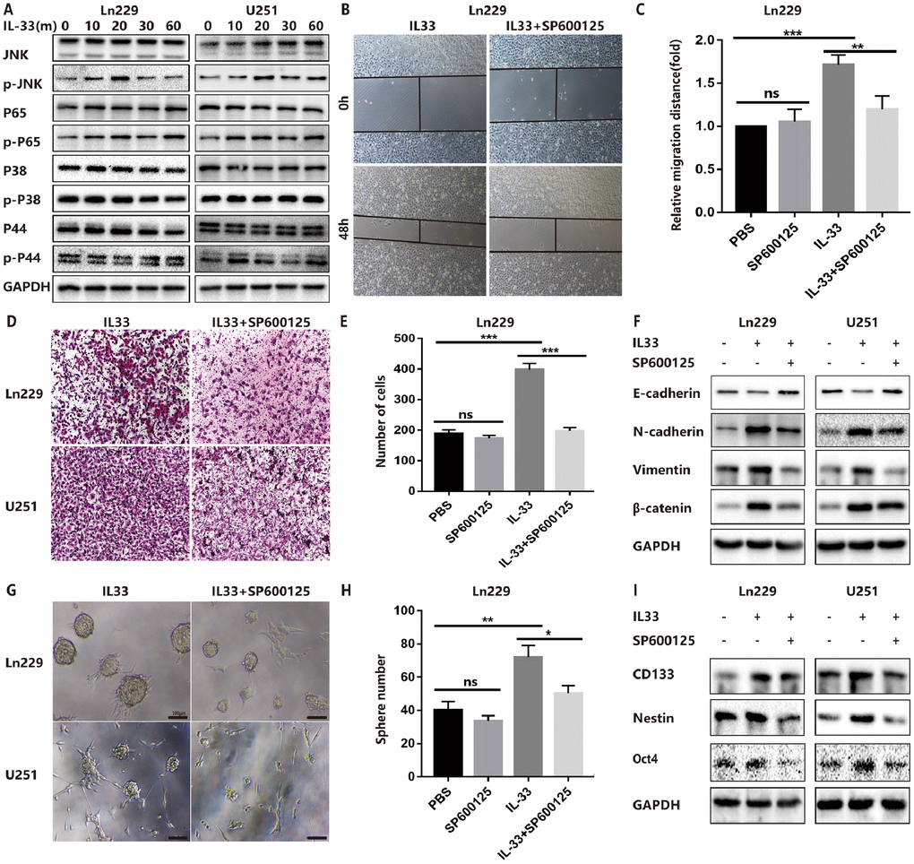
Figure 4.IL-33 promotes glioma EMT and stemness via JNK activation. (A) Effects of IL-33 on NF-κB and MAPK family signal in glioma cells. The cells were treated with IL-33 (20 ng/mL) in different periods of time. The expression of phosphorylated and total proteins was detected by Western blot. (B) Effects of the JNK inhibitor SP600125 on IL-33 induced migration by Wound healing assay. The cells were treated with IL-33 (20 ng/mL) and/or SP600125 (10 μg/mL) for 48 hours. (C) The tumor cells moving distance was detected. Experimental group (IL-33, SP600125 and IL-33+SP600125)/Control group were calculated for statistical analysis. Results are expressed as the mean ± SD; n=3; **, P < 0.01; ***, P < 0.001. (D) Transwell assay indicated the effects of SP600125 on IL-33 induced invasion. (E) The cells moved through the chambers from four groups (PBS, SP600125, IL-33 and IL-33+SP600125) were counted and analyzed. Results are expressed as the mean ± SD; n=3; ***, P < 0.001. (F) Effects of the JNK inhibitor SP600125 on EMT related proteins in glioma cells. N-cadherin, E-cadherin, Vimentin and β-catenin proteins were detected by Western blotting. (G) Effects of JNK inhibitor on glioma cells stemness. Glioma cell lines were subject to sphere formation assay. Representative images of spheres of glioma cells are shown. Scale bar, 100 μm. (H) Sphere number from four groups were counted and analyzed. Results are expressed as the mean ± SD; n=3; *, P < 0.05; **, P < 0.01. (I) The expression of CD133, Nestin and Oct4 were detected by Western blot.