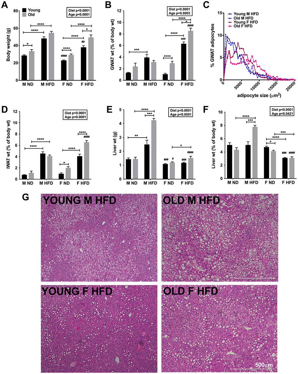
Figure 1.Aging and HFD feeding induced sex differences in total body adiposity and tissue weights. (A) Body weights of C57Bl6/j male and female on ND or 60% HFD starting at 6-week of age (young) or after 10 months of age (old) for 24-week. (B) GWAT percent weight. (C) Relative distribution of GWAT adipocyte cross-sectional area (D) IWAT percent weight. (E) Liver weight. (F) Liver percent weight. (G) H&E staining of liver sections depicting lipid accumulation in young and old obese male and females. Scale bar = 500 μm. N=7-12 /group. Two-way ANOVA with Bonferroni-Dunn’s post-test was performed for (A, B) and (D–F). Statistics from diet and sex interaction are in box. One-way ANOVA with Sidak’s post-test was performed for (C). Statistical significance is indicated by *p<0.05, **p<0.01, ***p<0.001, ****p<0.0001. Student’s t-test was performed for male and female comparisons between the same diet groups indicated by #p<0.05, ##p<0.01 ###p< 0.001 and ####p<0.0001; error bars are SEM.