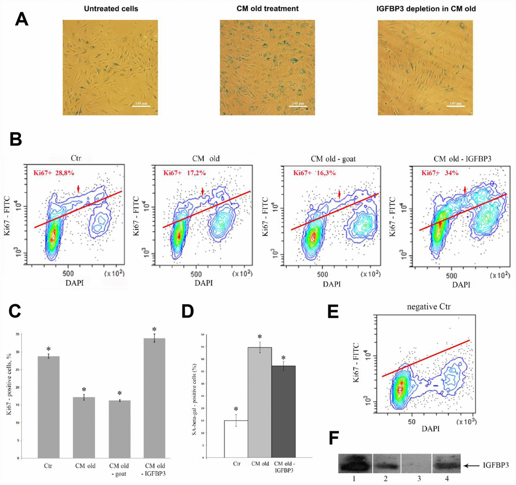
Figure 4.Effects of the IGFBP3-depleted CM old on young MESCs. (A, D) IGFBP3 immunodepletion decreases a population of SA-β-Gal positive cells. (A) SA-β-Gal staining 9 days after CM old treatment (middle) or after the IGFBP3-depleted CM old treatment (right). Representative microphotographs are shown. Ob: 10x; scale bars: 140 μm. (D) Quantitative assay of SA-β-Gal positive cells. Data are presented as mean ± SD, *p < 0.05. (B, C, E) The IGFBP3 immunodepletion increases the proliferation of young MESCs. (B) Ki67 staining. Cells were treated with indicated CMs for 9 days, stained with FITC-Ki67 conjugate and DAPI, and analyzed by FACS. (E) The negative control (negative Ctr) - FITC Mouse IgG1 staining. The representative FACS contour-plots of stained MESCs are shown; red arrows indicate S-phase cells. (C) Quantitative assessment of Ki67 positive cells. Ctr – untreated cells. The means ± SD of three independent experiments are presented, *p < 0.05. (F) Testing of IGFBP3 content in CMs by immunoblotting. 1 – recombinant IGFBP3, positive control; 2 – CM old after immunoprecipitation with normal goat IgG control antibodies, negative control; 3 – CM old after immunoprecipitation with specific IGFBP3 antibodies (IGFBP3-depleted CM old); 4 – CM from H2O2-treated (senescent) cells (CM old).