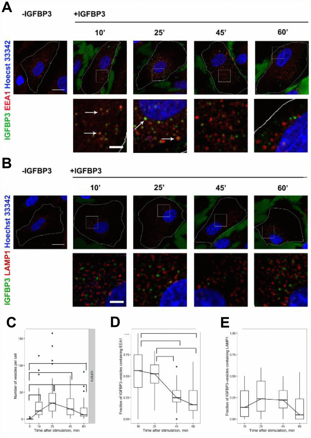
Figure 6.The analysis of exogenous rIGFBP3 colocalization with EEA1-positive early endosomes and LAMP1-positive late endosome/lysosomes. Exogenous rIGFBP3 was added to young MESCs for 10 min as indicated in Materials and Methods. Then the IGFBP3-treated cells (+IGFBP3) as well as control cells (-IGFBP3) were fixed and stained for IGFBP3 antibodies (green), EEA1 (A, red) or LAMP1 (B, red) and Hoechst 33342 (blue). White arrows indicate structures with the colocalization of IGFBP3 and endosome marker signals. Images are presented as the maximal intensity projections of three consecutive optical slices. Scale bars: 20 μm (upper panels) and 5 μm (lower panels). On the base of immunofluorescent images, the number of IGFBP3 vesicles (C) as well as the fraction of IGFBP3 vesicles containing EEA1 (D) or LAMP1 (E) were calculated. Data are presented as boxplots with median, interquartile range, minimum and maximum. Parentheses indicate statistical differences (p<0.05).