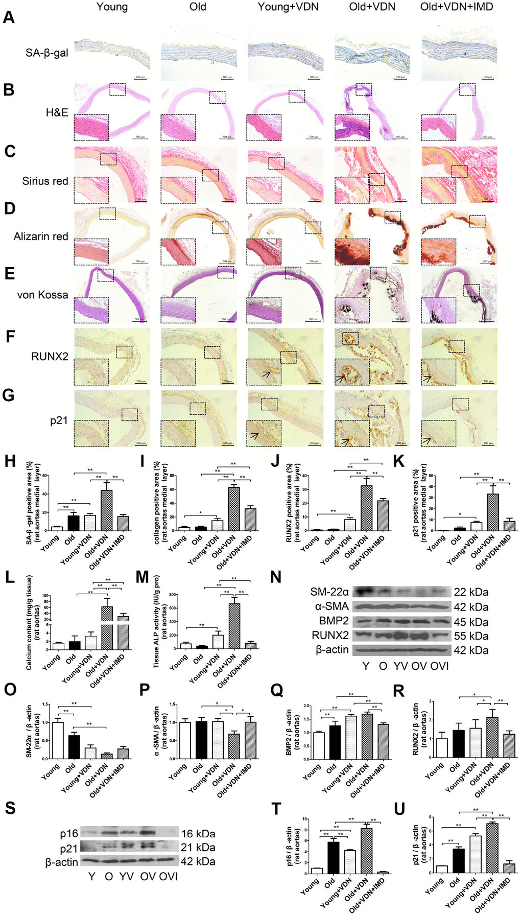
Figure 2.Exogenous IMD1-53 attenuated aging-associated vascular calcification in rats. (A) SA-β-gal staining for β-galactosidase activity (positive staining: blue) (Scale bar=100 μm), (B) H&E staining (Scale bar=500 μm), (C) Sirius red staining for collagen (red) (Scale bar=200 μm), (D) Alizarin red staining (red) (Scale bar=500 μm) and (E) von Kossa staining for vascular calcium deposition (black) (Scale bar=500 μm), (F) Immunohistochemistry staining for runt-related transcription factor 2 (RUNX2) (Scale bar=200 μm) and (G) cyclin-dependent kinase inhibitor p21 (Scale bar=200 μm), and (H–K) quantification of (H) β-galactosidase-positive staining (n=4), (I) collagen-positive staining (n=4), (J) RUNX2-positive staining (n=4), and (K) p21-positive staining (n=4) in the medial layer of rat thoracic aortas. (L) Calcium content assay (n=6) and (M) alkaline phosphatase (ALP) activity assay (n=6) in rat aortas. (N) Western blot analysis of protein levels of smooth muscle 22 alpha (SM-22α), alpha smooth muscle actin (α-SMA), bone morphogenetic protein 2 (BMP2) and RUNX2 in rat aortas, and (O–R) quantification (n=3). (S) Western blot analysis of protein levels of p16 and p21 in rat aortas, and (T, U) quantification (n=3). Enlarged regions, ×400. The arrow indicates positive staining. Y=young rats; O=old rats; YV=young+VDN; OV=old+VDN; OVI=old+VDN+IMD1-53. Data are mean ± SD. *P<0.05, **P<0.01.