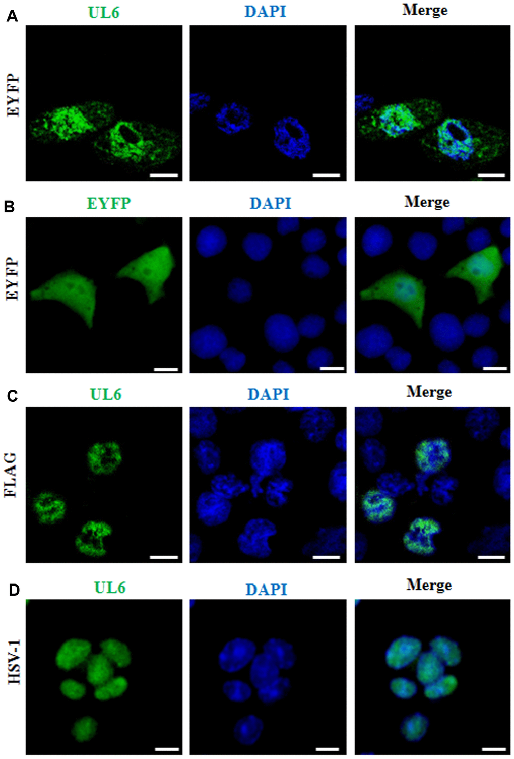
Figure 1.Subcellular distribution of UL6 in plasmid-transfected and HSV-1-infected cells. Subcellular distribution of EYFP-UL6 (A), EYFP (B) and FLAG-UL6 (C) in related plasmid transfected COS-7 cells. (D) Subcellular distribution of UL6 in HSV-1 infected Vero cells. Vero cells were infected with HSV-1 (F strain) at an MOI of 1. 8 h post-infection, Vero cells were fixed with 4% paraformaldehyde, permeabilized with 0.5% Triton X-100, and incubated with the anti-UL6 pAb. Then, cells were incubated with FITC-conjugated goat anti-rabbit IgG (green) and stained with DAPI (blue) to visualize the nuclei. EYFP fusion proteins were shown in pseudocolor green. The image shown represents a great proportion of the cells with homogeneous subcellular distribution. All scale bars indicate 10 um. Statistical analysis of the fluorescence was shown in Table 1.