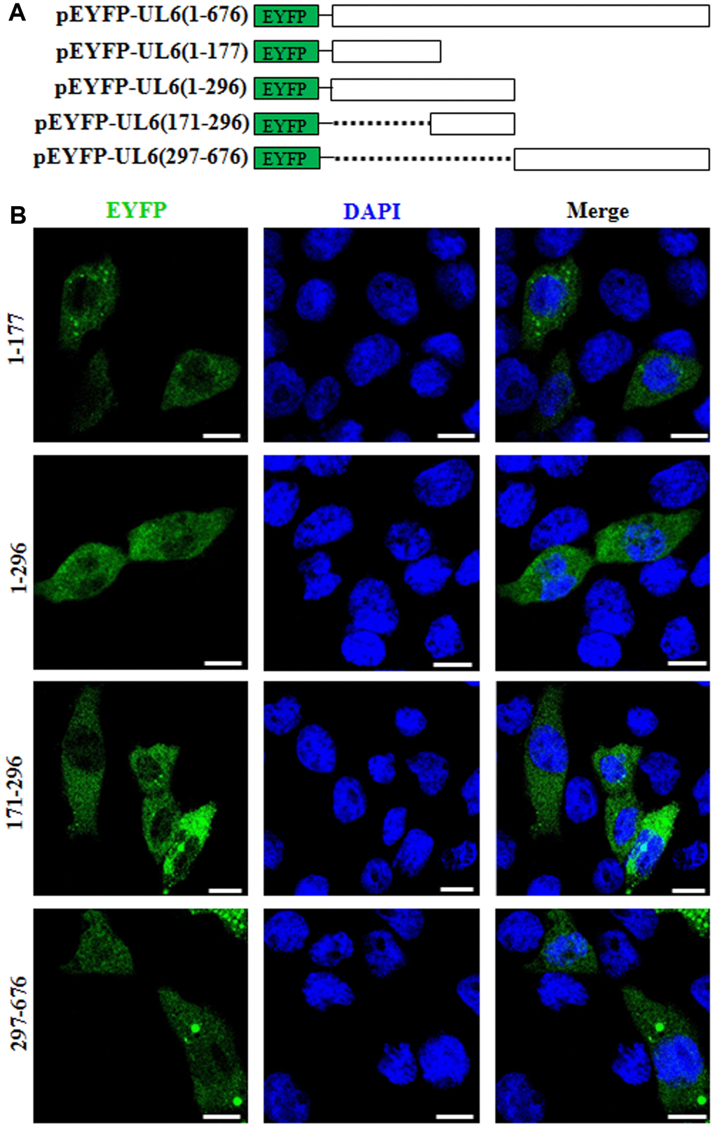
Figure 2.Subcellular distribution of the UL6 deletion mutants. (A) Schematic representation of wild-type UL6 protein and its N- and C-terminus deletion mutants fused with the C-terminus of EYFP. (B) Subcellular distribution of these UL6 deletion mutants shown in (A). Cells were stained with DAPI to visualize the nuclei. All scale bars indicate 10 um. Statistical analysis of the fluorescence was shown in Table 2.
Figure 2 — Molecular anatomy of the subcellular localization and nuclear import mechanism of herpes simplex virus 1 UL6 | Aging