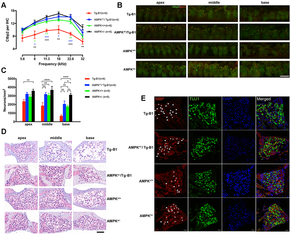
Figure 3.Downregulation of AMPK protects IHC ribbon synapses and SGNs. (A) Aging Tg-B1 mice showed reduced number of IHC synapses across all frequencies ranged from 5.6 to 32 kHz (F(1,10)=25.6, p=0.0005, two-way ANOVA followed by Bonferroni post-test) as compared to wild type controls. Significant increases of ribbon counts in IHCs at 8, 11.3, 16 and 22.6 kHz regions were observed in AMPK+/−/Tg-B1 (blue) mice compared to Tg-B1 (red) mice (F(1,10)=34.23, p=0.0002, two-way ANOVA followed by Bonferroni post-test) and the former showed almost similar numbers of ribbons to that in WT controls (green). Data are presented as the mean ± SEM, * P<0.05, ** P<0.01, ***P<0.001. n=6. (B) Representative z-stack confocal images in the IHC synapse areas from apical, middle and basal cochlear turns in four genotype groups aged 10-12 months showed co-staining in cochlear whole mount preparations with the presynaptic (CtBP2 for RIBEYE, Green puncta) and postsynaptic marker (GluR2, red puncta). CtBP2 in the IHC areas, seen as a cloud of ~0.4-0.6 um puncta, clustered at the basolateral pole. The IHC nuclei were also labeled due to the nuclear expression of CtBP2. Scale Bar=10 μm. (C) Statistics of SGN density (Number of SGNs/Area of Rosenthal’s canal) showing the significant SGNs degeneration in Tg-B1 (red bars) mice as compared to WT controls (green bars) (F(1,10) =40.67, p<0.0001, two-way ANOVA followed by Bonferroni post-test), especially in the middle (p=0.0016) and basal turns (p=0.0045) of the cochleae. SGNs survival in AMPK+/−/Tg-B1 mice (blue bars) has a remarkable increase compared to Tg-B1 mice (F(1,10)=59.99, p<0.0001, two-way ANOVA followed by Bonferroni post-test), especially in the middle (p=0.0017) and basal turns (p=0.0011) of cochleae. (D) Representative H&E staining images of SGNs taken from cochleae of aging mice at 10-12 months. The neurons in AMPK KO mice were arranged tightly whereas significant reduction of SGN number occurred in the basal turn of WT controls and more aggravated in the middle and basal turns in Tg-B1 mice. Scale bar=50 μm. (E) Representative immunostaining for MBP expression in SGN in four genotype mice. MBP+ myelin sheaths (red) enclose type I SGNs (green) in Rosenthal’s canal of the middle turn of the cochlea. SGNs are co-identified with DAPI (blue) and TUJ1(green) staining. The MBP+ myelin sheath was considered intact if enveloped more than 80% of the outline of the perikarya. Intact MBP+ myelin sheaths are marked by asterisks while broken MBP+ myelin sheaths are indicated by arrows. A decline of intact MBP+ myelin sheath was found in Tg-B1 mice cochlea. Scale bar=20 μm.