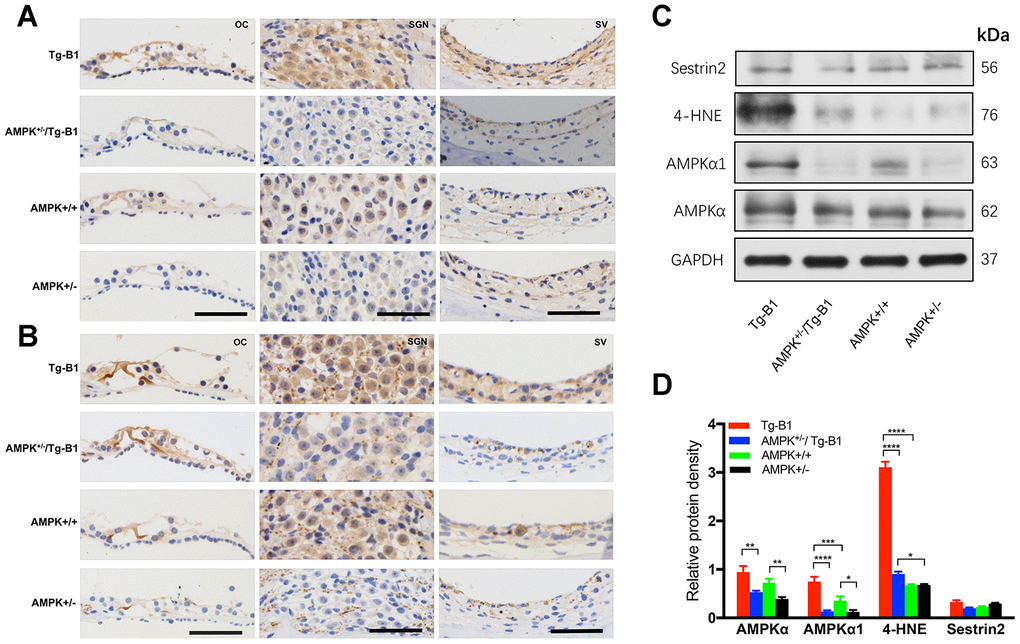
Figure 6.Expression of AMPK and p-AMPK, quantification of ROS and antioxidant protein in the cochlear tissues. (A–B) The representative mid-modiolar immunohistochemistry staining of cross-sections of the cochlea for the expressions of total AMPKα1 and p-AMPKα in three regions of the cochlea: OC (left column), SGN (middle column) and SV (right column). Increased DAB-stained immunolabeling of AMPKα1 and p-AMPKα (brown) in the cytosol and nuclei of OHCs, IHCs, OC, SGNs, basal cells of the SV were observed in the cochlear sections of Tg-B1 mice than those in AMPK+/−/Tg-B1 and WT mice. There was strong immunolabeling for p-AMPKα in the OC, SGNs, and SV of Tg-B1 mice, while AMPK+/− showed the weakest immunolabeling signals. Scale bar=50 μm. (C) Western blot using sensory epithelium tissues from 10-12 months mice displayed significant alteration in band density for total AMPKα, AMPKα1, and 4-HNE in the cochleae between Tg-B1 and AMPK+/−/Tg-B1 mice, but no significant difference in Sestrin2 expression, the antioxidant protein. GAPDH served as the loading control. (D) Histograms (mean ± SEM) represent relative density values normalized to GAPDH. Blotting results of AMPKα1 showed knockouts of AMPKα1 in Tg-B1 mice significantly decreased the AMPKα1 (p<0.0001) expression in the inner ear. Western blot analyses of 4-HNE expression in Tg-B1 mice cochleae were significantly higher than WT controls (p<0.0001) and KO mice (p<0.0001). Experiments were performed in triplicate, and p-values were determined by one-way ANOVA followed by Bonferroni post-test.