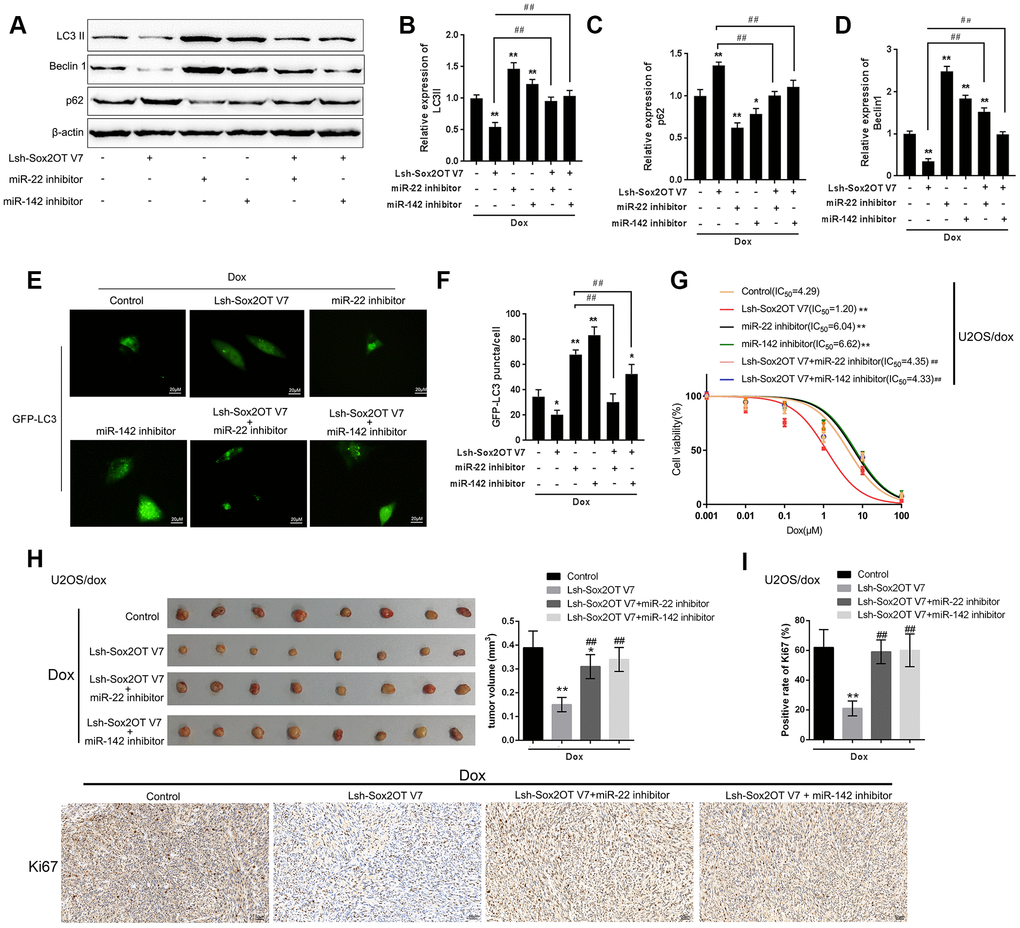
Figure 6.Dynamic effect of Sox2OT-V7, miR-142, and miR-22 on Dox-induced autophagy in U2OS cells. (A–D) OS cells were c-transfected with Lsh-Sox2OT-V7 and miR-142 inhibitor or miR-22 inhibitor upon Dox treatment and examined for the protein levels of LC3 II, Beclin 1, and p62 were examined. (E, F) U2OS cells with stable eGFP-LC3 expression were cotransfected with Lsh-Sox2OT-V7 and miR-142 inhibitor or miR-22 inhibitor, treated with Dox (5 μM) for 24 h, and examined for the formation of puncta by using a confocal microscope. Representative images are presented. (G) U2OS/Dox cells were assigned to six groups: control group, single Lsh-Sox2OT-V7 group, single miR-22 inhibitor group, single miR-142 inhibitor group, Lsh-Sox2OT-V7 + miR-22 inhibitor group, and Lsh-Sox2OT-V7 + miR-142 inhibitor group. Cells were transfected accordingly, treated with a series of concentrations of Dox (0.001, 0,01, 0.1, 1, 10, and 100 μM), and examined for the cell viability by MTT assay. (H) An in vivo tumor xenograft assay was performed by injecting U2OS/Dox cells that were not infected, infected with Lsh-Sox2OT-V7, transduced with Lsh-Sox2OT-V7 + miR-22 inhibitor, or transduced with Lsh-Sox2OT-V7 + miR-142 inhibitor under Dox treatment (n = 8 in each group). Tumors were shown and tumor volumes were detected. (I) Cell proliferation within the tumor was determined by IHC staining using an anti-Ki67 antibody. The data are presented as the mean ± SD of three independent experiments. *P<0.05, **P<0.01, compared to the control group; #P<0.05, ##P<0.01, compared to the Lsh-Sox2OT-V7 group.