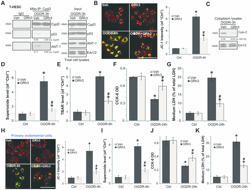
Figure 2.GRh3 protects endometrial cells from OGDR. T-HESC cells (A–G) or the primary murine endometrial cells (H-K) were pre-treated with GRh3 (10 μM, 2h pretreatment), followed by OGDR stimulation. After indicated time periods, mitochondrial CypD-p53-ANT-1 association (“Mito-IP”, A), mitochondrial depolarization (the JC-1 green intensity, B and H), cytochrome C (“Cyto-C”) release (C, testing the cytosol proteins), as well as superoxide contents (D and I) and lipid peroxidation (TBAR assay, E) were tested; Cell viability and necrosis were tested by CCK-8 (F and J) and medium LDH release (G and K) assays, respectively. For the JC-1 assays the representative JC-1 images, integrating both green and red fluorescence images, were presented (same for all Figures). For the Mito-IP assay, CypD-bound p53 and ANT-1 were quantified (A). For the cytochrome C release measurement, cytosol cytochrome C (vs. Tubulin) was quantified (C). Error bars stand for mean ± standard deviation (SD, n=5). “Ctrl” stands for “Mock” control treatment. *p<0.05 vs. “Ctrl”. #p<0.05 vs. cells with “Veh” pretreatment. Each experiment was repeated three times with similar results obtained. Bar=100 μm (B and H).