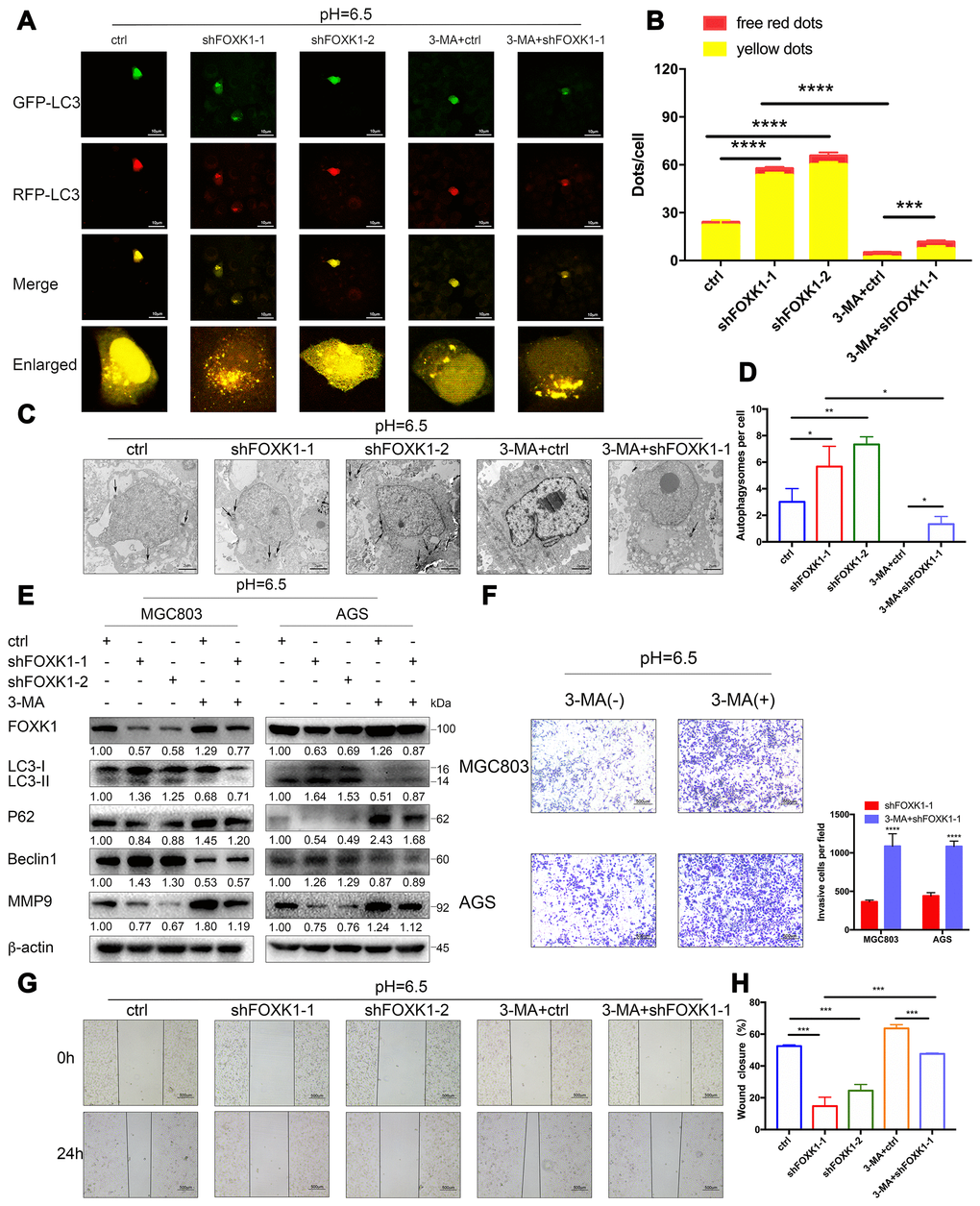
Figure 4.The silencing of FOXK1 induces autophagy to inhibit the migration and invasion of acidic GC cells. (A–H) Acidic MGC803 and AGS cells were infected with an empty lentivirus control (LV-ctrl) or with LV shFOXK1-1 or shFOXK1-2 and subsequently incubated for 24 h prior to pretreatment with 2 mM 3-MA or PBS (control). (A, B) Laser confocal microscopy analysis and quantification of transfected MGC803 cells with plasmid constructs containing LC3, which was fused with tandem mRFP-GFP tags. Scale bar, 10 μm. (C, D) The number of autophagosomes was observed and quantified under a transmission electron microscope. Scale bar, 2 μm. (E) Western blotting analysis of the levels of FOXK1, LC3-I, LC3-II, P62, Beclin1 and MMP9; β-actin was used as a loading control. (F) Matrigel cell invasion assays of MGC803 and AGS cells were performed, and the invading cells were quantified. (G, H) Scratch test evaluation of MGC803 cells. The wound healing area was analyzed using ImageJ software. Scale bar, 500 μm. The data are presented as the means ± S.D.s from three independent experiments. * P < 0.05, ** P < 0.01, *** P < 0.001, and **** P < 0.0001.