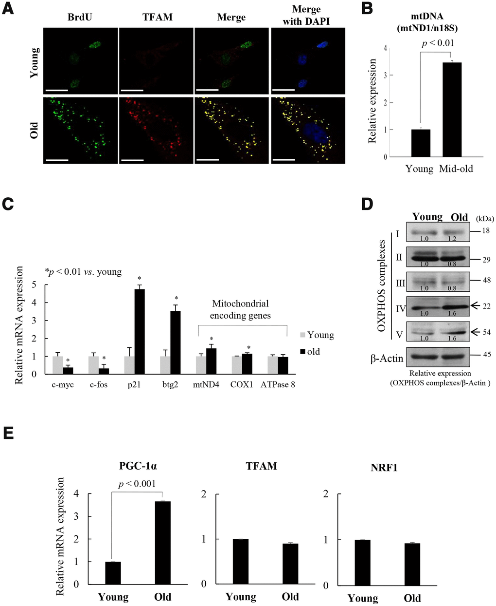
Figure 1.Replicative senescence of foreskin human diploid fibroblast (fs-HDF) is accompanied by mitochondrial nucleoid remodeling and biogenesis. (A) Immunofluorescence staining of BrdU incorporation into young and old fs-HDF cells. BrdU (green) and mitochondrial transcription factor A (TFAM; red) were observed by confocal microscopy. Nuclei were stained with DAPI (blue). Scale bars, 25 μm. (B) RT-qPCR analysis of the mitochondrial DNA (mtDNA) level, normalized to that of nDNA. (C) RT-qPCR analysis of the expression of nuclear- and mitochondrial-encoded genes. The expression levels of mitochondrial OXPHOS complex-I, -IV, and -V; c-myc and c-fos (markers of proliferation), and p21WAF1 and btg2 (markers of cell-cycle arrest) were analyzed. (D) Immunoblot analysis of OXPHOS complex I–V. β-actin was used as the loading control. Band intensity was quantified using ImageJ software (NIH, Bethesda, MD, USA) and normalized to that of β-actin. (E) Relative mRNA levels of PGC-1α, TFAM, and NRF1 by RT-qPCR. Data are means ± standard deviation (SD) of three independent experiments per group.