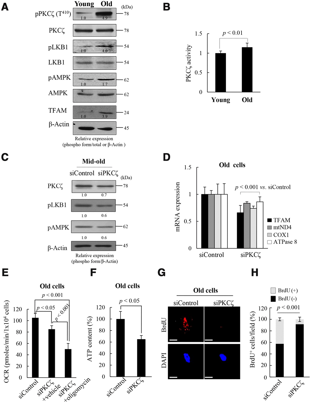
Figure 2.Protein kinase C zeta (PKCζ) regulates mitochondrial reprogramming in old fs-HDF cells. (A) Immunoblot of PKCζ, LKB1, AMPK, and TFAM in young and old cells. (B) PKCζ was purified from young and old cells by immunoprecipitation (IP) and subjected to in vitro kinase assay using a PKC kit. (C) Mid-old fs-HDF cells were transfected with siRNAs-PKCζ and subjected to immunoblot analysis. Band intensity was quantified using ImageJ software and normalized to β-actin. (D) Old fs-HDF cells transfected with siPKCζ were subjected to RT-qPCR to measure the expression of TFAM and the mitochondrial complex-I (ND4), -IV (COX1), and -V (ATPase8) subunits. Data were normalized to the siControl-transfected cells. (E) Old fs-HDF cells transfected with siPKCζ were treated with or without oligomycin (10 μM) for 1 h and the oxygen consumption rate (OCR; pmol/min/1 × 104 cells) was compared to that of siControl-transfected cells. Note significant inhibition of OCR by knockdown of PKCζ expression. It was more downregulated by oligomycin cotreatment. (F) ATP levels in old cells transfected with siPKCζ. (G) BrdU incorporation in old cells transfected with siControl or siPKCζ. Nuclei were stained with DAPI (blue). Scale bars, 10 μm. (H) BrdU incorporation in mitochondria was quantified. Confocal microscope images were captured and counted at least 200 cells using ImageJ software (n=10 images/group). Data are means ± SD of two independent experiments per group. Student’s t-test or one-way ANOVA followed by Tukey HSD post hoc test.