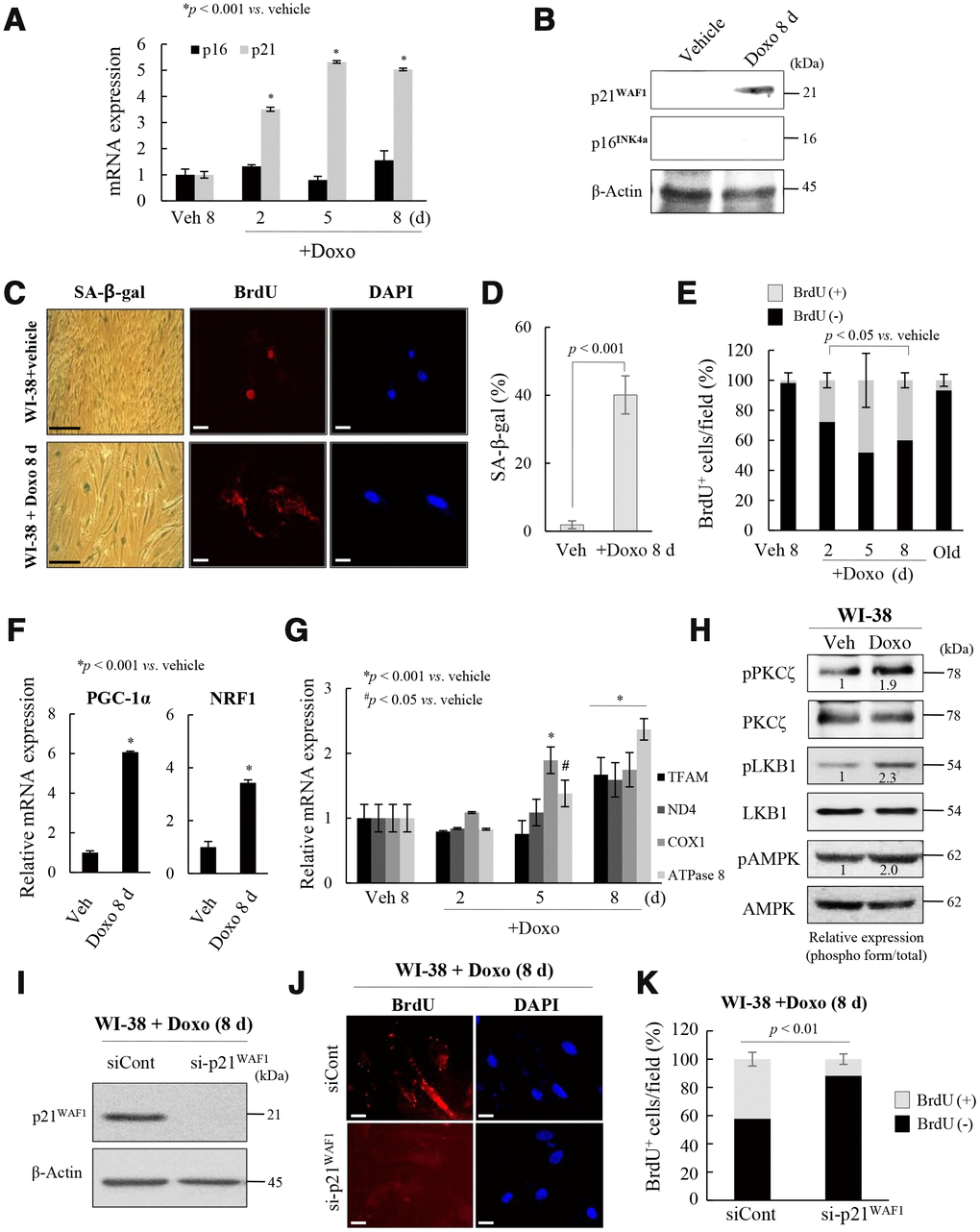
Figure 4.Activation of p21WAF1regulates mitochondrial reprogramming in senescent WI-38 cells. (A) Young WI-38 cells were treated with DOXO (100 ng/mL) for 24 h and maintained for 8 days; the expression of p21WAF1 and p16INK4a was assayed by RT-PCR at the indicated times. (B) Immunoblot analysis. Note induction of p21WAF1, but not p16INJ4a, expression in young cells after Doxo treatment. (C) SA-β-galactosidase expression and BrdU incorporation in mitochondria in young cells after Doxo treatment for 8 days. Nuclei were stained with DAPI. Scale bars, 100 μm (black bar) or 20 μm (white bar). (D) BrdU incorporation in mitochondria was quantified. More than 300 cells were counted in 5 fields. (E) Cells with BrdU (+) mitochondria were counted under a confocal microscope using ImageJ software (n = 20 images/group). (F) RT-qPCR analysis of PGC-1α and NRF1 expression in Doxo-treated WI-38 young cells. (G) The expression of TFAM and the complex-I, -IV, and -V subunits in Doxo-induced senescent WI-38 cells was measured by RT-qPCR. (H) Immunoblot analysis reveals activation of PKCζ, LKB1 and AMPK after Doxo treatment. Band intensity was quantified using ImageJ software. (I) Doxo treated WI-38 cells were transfected with siRNAs-p21WAF1 and subjected to immunoblot analysis. (J) Immunocytochemistry showing the loss of BrdU incorporation in mitochondria by knockdown of p21WAF1 expression in Doxo-treated cells. Nuclei were stained with DAPI. Scale bars, 20 μm. (K) BrdU incorporation in mitochondria was quantified. Confocal microscope images were captured and counted at least 200 cells using ImageJ software (n=10 images/group). Data are means ± SD of three independent experiments per group.