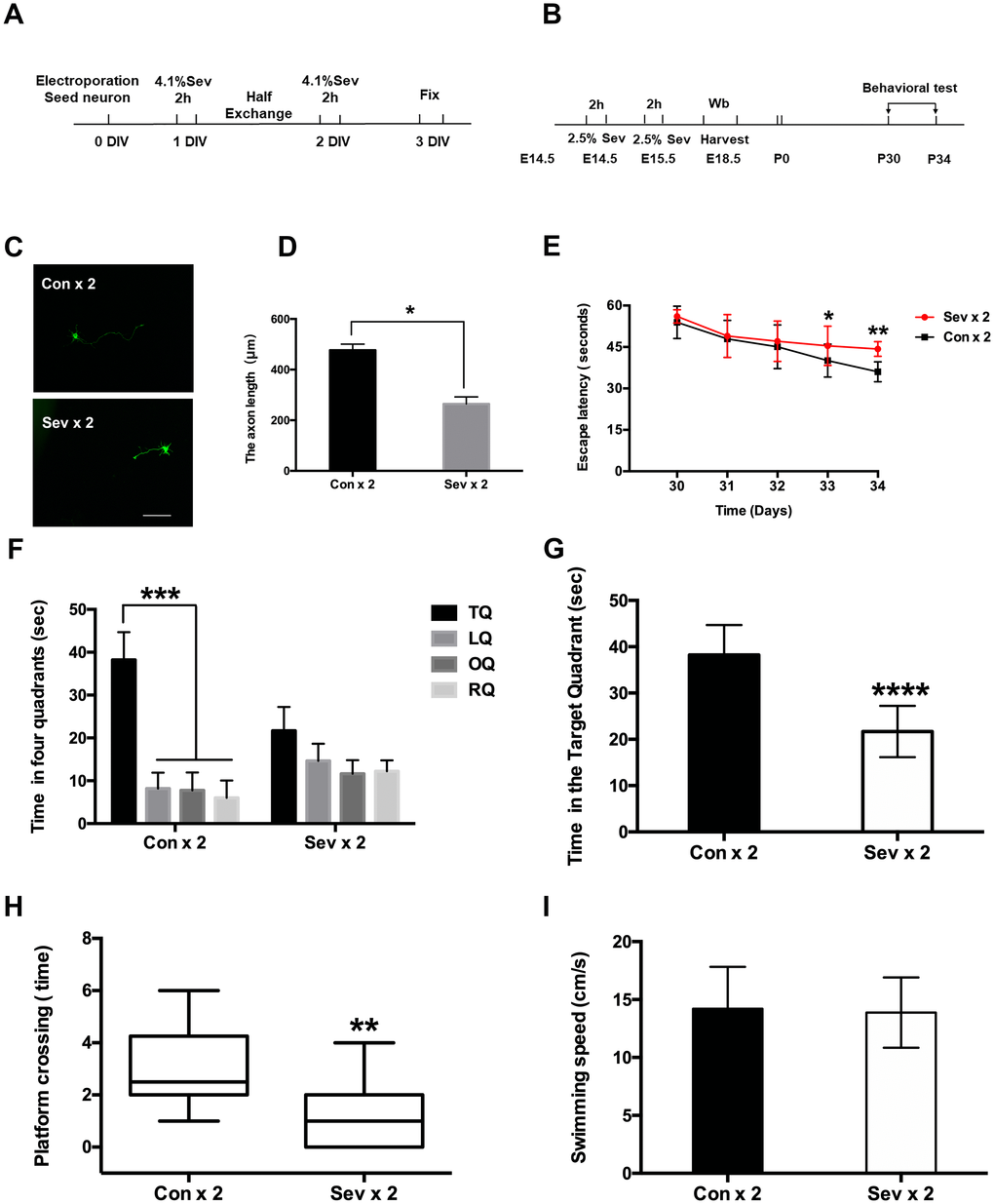
Figure 1.Effects of sevoflurane anesthesia on spatial learning and memory in young mice. (A) Flowchart of the neuron electroporation experiment. (B) Flowchart of the MWM experiment. (C) Dual sevoflurane exposure decreased axon length in primary cultured mouse cortical neurons. (D) The statistical results for the axon length between the two groups. Scale bars = 100 μm; approximately 70 cells from three independent experiments were counted during the statistical analysis (P = 0.0147*, Student’s t-test). (E) The escape latency on the 4th day of acquisition training was increased in the sevoflurane group (Sev x 2 vs Con x 2, F = 0.828, P = 0.028*, Student’s t-test, N = 10). During the probe trial, the escape latency was also increased in the dual sevoflurane group (Sev x 2 vs Con x 2, F = 1.35, P = 0.007**, Student’s t-test, N = 10). (F) During the probe trial, the control group spent much more time in the target quadrant than other quadrants (P < 0.001***, N = 10, one-way ANOVA), while the sevoflurane group spent similar periods in the four quadrants (P > 0.05, N = 10, one-way ANOVA). TQ, LQ, OQ, and RQ is the target quadrant, the left quadrant, the opposite quadrant, and the right quadrant, respectively. (G) Dual sevoflurane exposure decreased the time spent in the target quadrant (F = 0.143, P < 0.0001****, N = 10, Student’s t-test). (H) Sevoflurane decreased the platform crossing times (F = 1.156, P = 0.0033**, N=10, Student’s t-test). (I) Sevoflurane did not affect swimming speed compared with the same variables in the control group mice. Data are expressed as the means ± S.D. *P < 0.05, **P<0.01, ***P<0.001.****P < 0.0001.