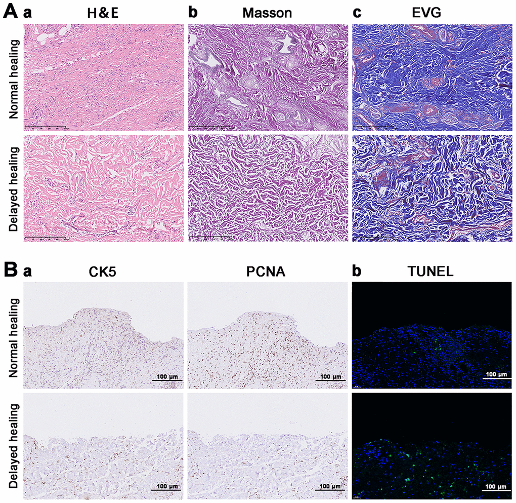
Figure 1.Impaired tissue remodeling and re-epithelization in delayed cutaneous wound tissue. (A) Histological staining of cutaneous wound tissue (200×). (a) H&E staining of sustained inflammatory cells and disordered tissue organization in the delayed cutaneous wound. (b) Masson staining of collagenous (blue) and muscular (red) fibers. Less staining was observed in the delayed wound tissue. (c) EVG staining of elastic fibers. Less staining occurred in the delayed wound tissue. (B) IHC and TUNEL staining of cutaneous wound tissue (200×). (a) CK-5 and PCNA expression levels are reduced in the delayed wound tissue. (b) Increased apoptosis (green cells) occurred in the delayed wound tissue. All the experiments were repeated at least three times.