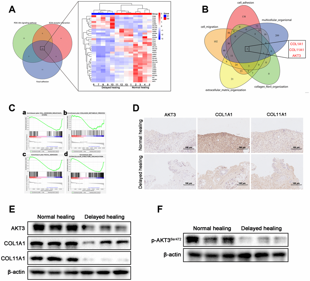
Figure 3.Downregulation of AKT3, COL1A1, and COL11A1 in delayed cutaneous wound tissue. (A) Venn diagram of the KEGG pathway. (a) Venn analysis identified 35 genes that were enriched in PI3K-AKT signaling, ECM-receptor interactions, and focal adhesion. (b) The heatmap expression profile of the 35 changed genes. (B) Venn diagram of GO analysis for the tissue remodeling-associated biological functions. AKT3, COL1A1, and COL11A1 were enriched. (Ca–d) Gene set enrichment analysis (GSEA) of cutaneous wound tissue. The genes associated with (a) cell adhesion molecules, (b) collagen metabolic processes, (c) focal adhesion, and (d) extracellular structural organization were negatively enriched in the delayed cutaneous wound tissue. (D) IHC staining of AKT3, COL1A1, and COL11A1 in cutaneous wound tissue (200 x). The levels of all three proteins were reduced in the delayed wound tissue. (E) Decreased AKT3, COL1A1, and COL11A1 protein levels in delayed cutaneous wound tissue. (F) Total AKT3 and phosphorylated-Ser472 AKT3 levels were decreased in delayed cutaneous wound tissue. All the experiments were repeated at least three times.