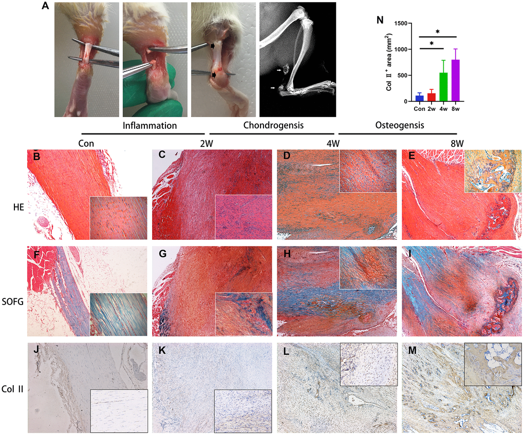
Figure 1.Endochondral ossification is the pathological basis of the heterotopic ossification model in the rat after tendon injury. (A) The HO model was made by complete transverse incision at the midpoint of Achilles tendon without any attempt of repair. Bone-like tissues formed near the position of the tendon-to-bone junction and tendon-to-muscle junction (arrow) at 8 weeks post-surgery. (B–I) The H&E and Safranin O/fast green staining. Inflammation infiltration was obvious at 2w, lots of chondrocytes could be found at 4w, and bone tissues formed at 8w. (J–N) The immunohistological staining of Col II + cells (brown). *p < 0.05 as determined by one-way ANOVA test.