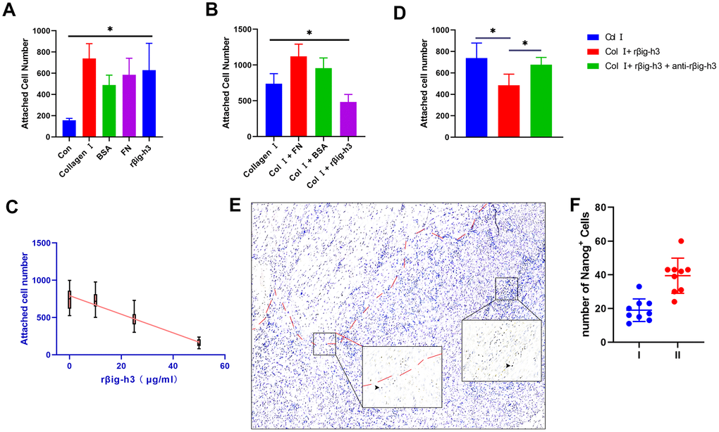
Figure 4.Cell attachment of βig-h3 to iTDSCs. (A) The cell attachment of iTDSCs to collagen I, BSA, FN, rβig-h3, or nothing. (B) iTDSCs attachment to collagen type I in the presence of BSA, FN, or rβig-h3. (C) The association between the dose of rβig-h3 and the number of attached cells. (D) The cell attachment of iTDSCs to collagen I in the presence of rβig-h3 with/without anti-rβig-h3. (E, F) The immunohistological staining of Nanog+ cells (brown, black arrow). The area above the red dotted line is the injured tendon. The positive cells were counted as the follows: I. area around the tendon (half of the 40X view is the tendon tissue) II. area except the tendon (without any tendon tissue)*p < 0.05 as determined by one-way ANOVA test.