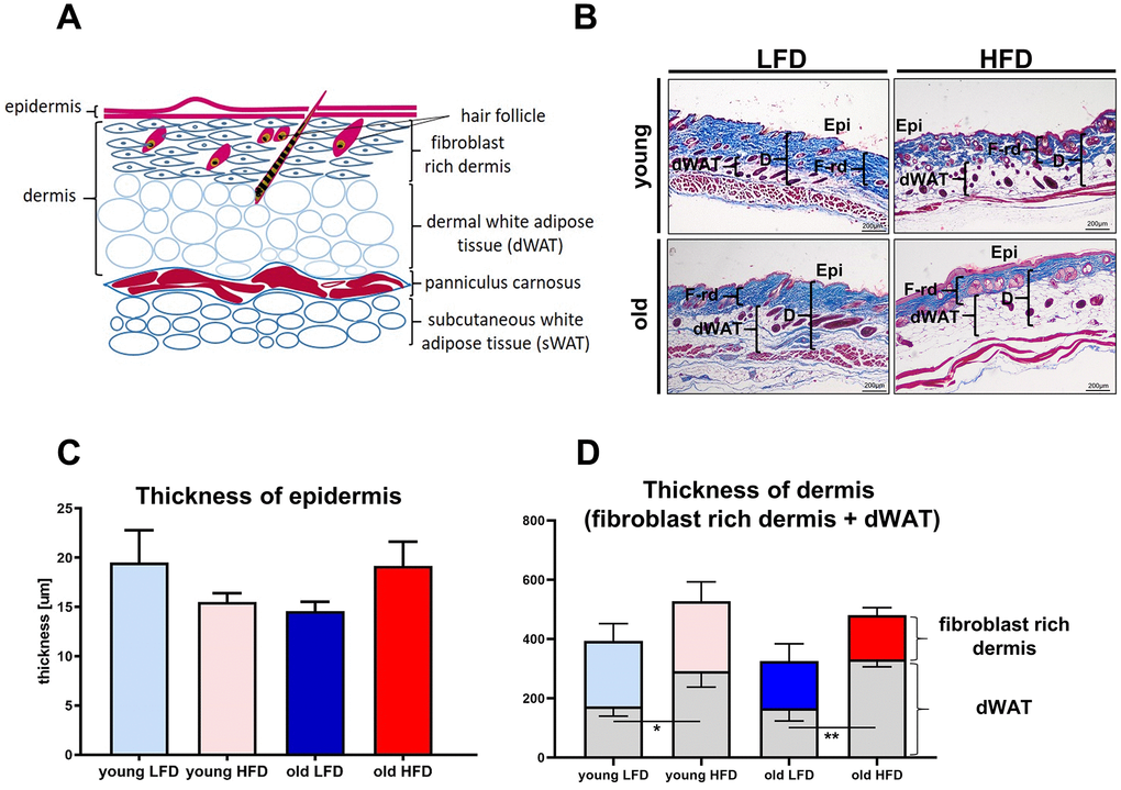
Figure 3.Histological analysis of skin structure and thickness. Scheme of skin structure (A), histological skin sections stained with Masson trichrome and collected from young or old mice fed for a period of 8 weeks LFD or HFD (B), quantification of the skin layers thickness: epidermis (C) and dermis (D). The measurement of skin thickness were performed on histological slides collected from n=24 mice (n=6 per group). Epi - epidermis, dWAT - dermal white adipose tissue, F-rd fibroblast rich dermis; scale bar 200 μm, The bars indicate lsmean ±SE *p<0.05, **p<0.01.