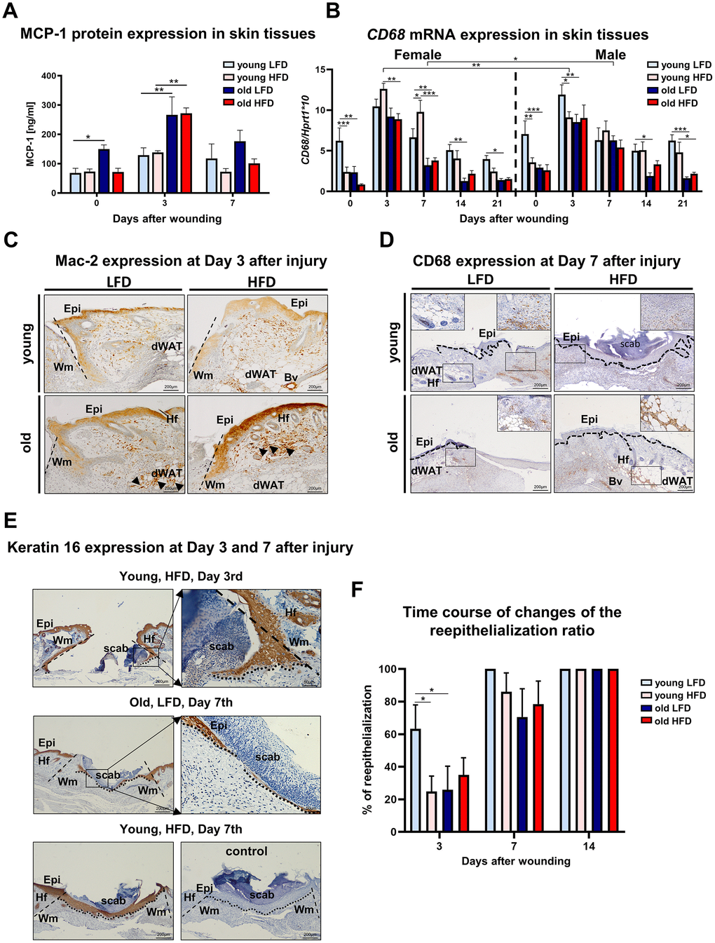
Figure 4.Inflammatory response and histological analysis of re-epithelialization during skin wound healing. (A) MCP-1 protein levels (n=6 skin samples per group); (B) CD68 mRNA expression (n=4-8 skin samples per group); (C) Mac-2 and (D) CD68 immunohistological localization on skin tissues at post-wounded day 3 (C) and day 7 (D). Immunohistochemical detection of keratin 16 (E) and morphometrical analysis (F) of the re-epithelization process in the skin of old, young, LFD or HFD mice (n = 3-5 mice per group). Epi - epidermis, dWAT - dermal white adipose tissue, Wm – wound margin, Hf – hair follicles; control (E) of immunohistochemical reaction where the primary antibody were omitted. Histological sections were counterstain with haematoxylin. Scale bar (C–E) 200μm, insets (C–E) 50μm. The bars indicate lsmean ±SE *p<0.05, **p<0.01, ***p<0.001.