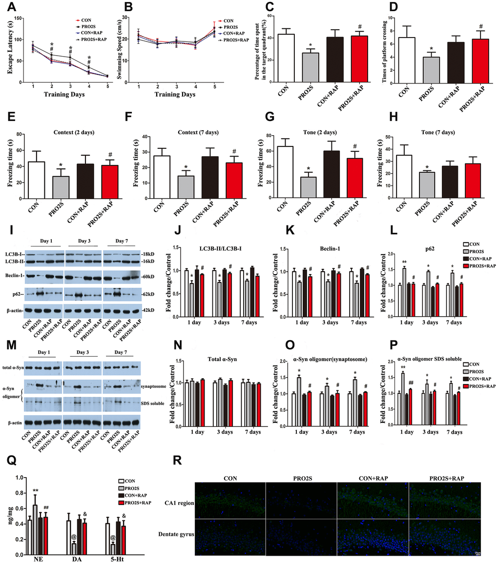
Figure 4.The effects of an autophagy agonist on changes in behavior, autophagy-related protein levels and α-synuclein levels induced by propofol anesthesia and surgery in aged rats. (A) Rapamycin (RAP) reversed the increased escape latency induced by propofol anesthesia (2 h) and surgery. (B) Propofol anesthesia (2 h) and surgery with or without rapamycin did not alter the swimming speed. (C, D) Rapamycin reversed the reduced target quadrant dwelling time and number of platform crossings induced by propofol anesthesia (2 h) and surgery. (E, F) Rapamycin ameliorated the reduced freezing time in the context test induced by propofol anesthesia (2 h) and surgery, both 2 and 7 days after surgery. (G, H) Rapamycin ameliorated the reduced freezing time in the tone test induced by propofol anesthesia (2 h) and surgery on day 2 but not day 7 after surgery. (I) Representative immunoblots illustrating the effects of rapamycin on the changes in hippocampal autophagy-related protein levels induced by propofol anesthesia (2 h) and surgery. (J) Rapamycin reversed the decrease in LC3B expression in the hippocampus induced by propofol anesthesia (2 h) and surgery. (K) Rapamycin reversed the decrease in Beclin-1 expression in the hippocampus induced by propofol anesthesia (2 h) and surgery. (L) Rapamycin reversed the increase in p62 expression in the hippocampus induced by propofol anesthesia (2 h) and surgery. (M) Representative immunoblots illustrating the effects of rapamycin on the changes in α-synuclein (α-Syn) levels in the hippocampus induced by propofol anesthesia (2 h) and surgery. (N) Rapamycin did not alter total α-synuclein expression. (O, P) Rapamycin reversed the α-synuclein oligomerization in synaptosomes and SDS-solubilized fractions in the hippocampus induced by propofol anesthesia (2 h) and surgery. (Q) Rapamycin reversed the changes in neurotransmitter levels in the hippocampus induced by propofol anesthesia (2 h) and surgery. (R) Representative confocal staining of LC3B expression in the CA1 and DG regions of the hippocampus in the four groups of rats 7 days after propofol anesthesia. Values are expressed as the mean ± SEM (n = 10 per group for behavioral tests, n = 8 per group for neurotransmitter detection, n = 6 per group for Western blot analysis, n = 4 per group for confocal analysis of LC3B staining). *p < 0.05, **p < 0.01, @p < 0.001 PRO2S vs. CON group. #p < 0.05, ##p < 0.01, &p < 0.001 PRO2S + RAP group vs. PRO2S group. CON: the control group, CON + RAP: the control + rapamycin group, PRO2S: the 2-h propofol anesthesia + surgery group, PRO2S + RAP: the 2-h propofol anesthesia + surgery + rapamycin group. Scale bar, 20 μm (R).