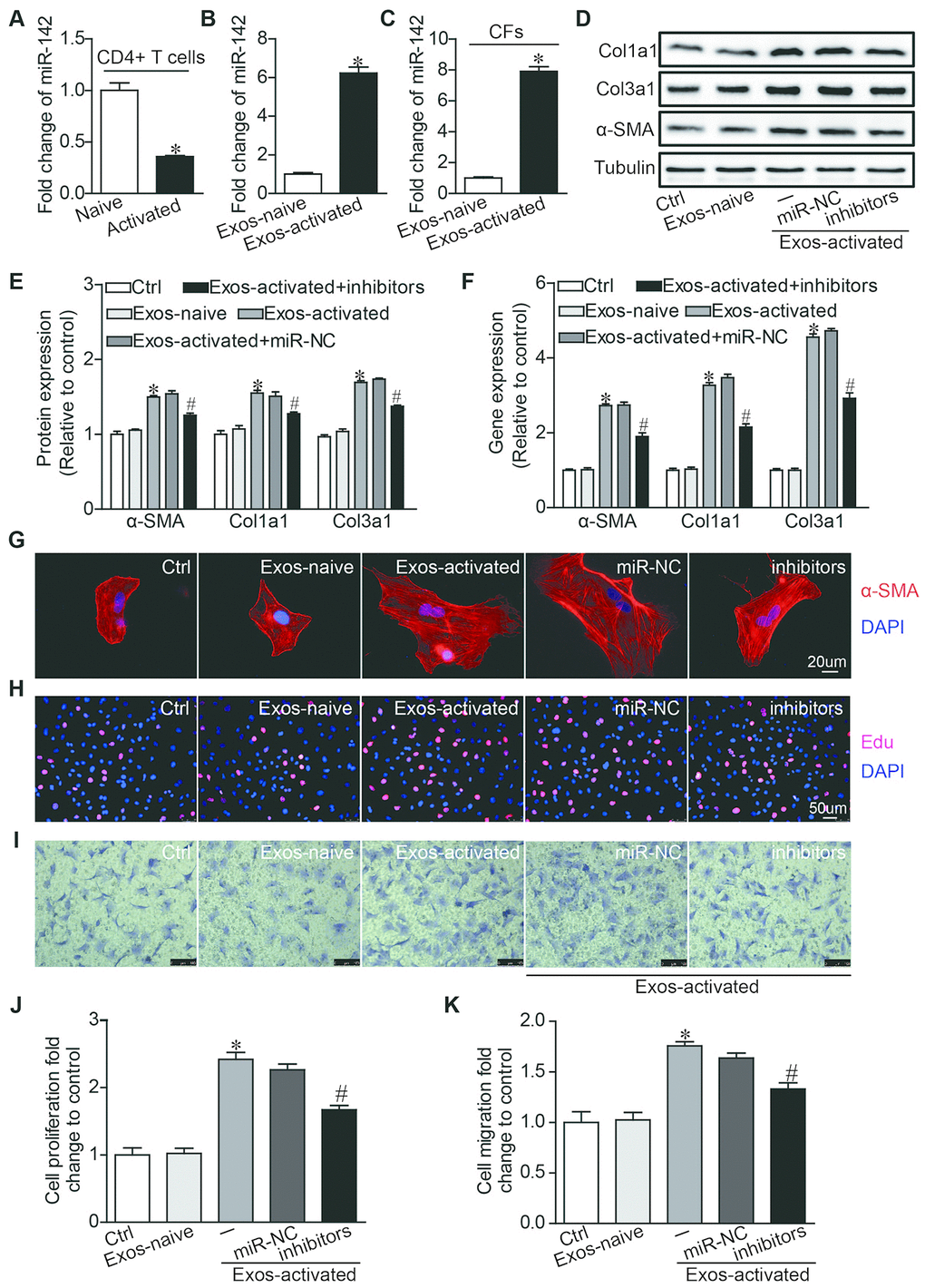
Figure 4.MiR-142 partially mediated the pro-fibrotic effects of activated CD4+ T cells-derived exosomes on cardiac fibroblasts. (A) MiR-142-3p expression was detected in naive CD4+ T cells and activated CD4+ T cells by qRT-PCR. n=3 per group. *P < .05. (B) MiR-142-3p expression was detected in exosome derived from naive and activated CD4+ T cells by qRT-PCR. n=3 per group. *P < .05. (C) MiR-142-3p expression was detected in CFs before and after incubated with exosomes derived from activated CD4+ T cells for 24h by qRT-PCR. n=3 per group. *P < .05. (D–F) Western blotting and qPCR analysis of α-SMA, Col1a1 and Col3a1 levels in cardiac fibroblasts. The blots shown are representative of three independent experiments. *P < .05 vs. Exos-naive. #p < .05 vs Exos-activated + miR-NC. (G) Immunofluorescent analysis of myofibroblast activation. The images shown are representative of three independent experiments. Red signals indicated α-SMA protein expression, and blue signals for nuclei. Scale bar = 20 μm. (H) Cardiac fibroblasts proliferation was detected using the EdU incorporation assay. The images shown are representative of three independent experiments. Scale bar = 50 μm. (I) Cardiac fibroblasts migration was detected using the transwell assay. The images shown are representative of three independent experiments. Scale bar = 100 μm. (J) Quantification analysis of cardiac fibroblasts proliferation using EdU assay data. *P < .05 vs. Exos-naive; #p < .05 vs. Exos-activated + miR-NC. (K) Quantification analysis of cardiac fibroblasts migration using Transwell assay data. *P < .05 vs. Exos-naive. #p < .05 vs. Exos-activated + miR-NC.