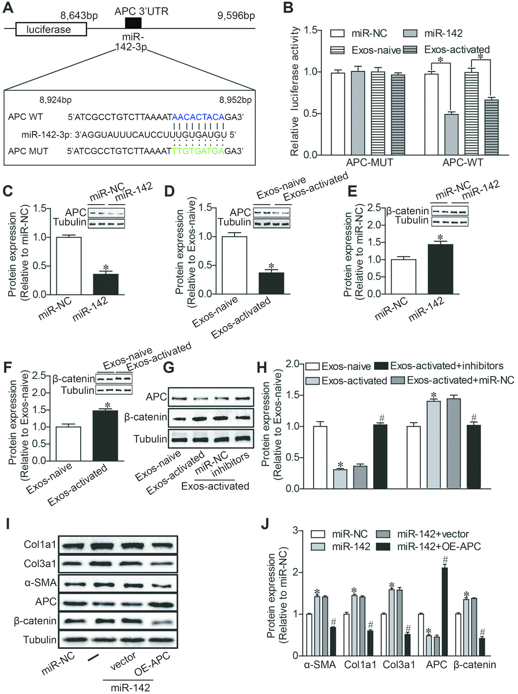
Figure 5.MiR-142 targets APC, resulting in the activation of WNT pathway. (A) Diagram of miR-142-3p binding site in APC 3′UTR. (B) Luciferase reporter assay of the interaction between miR-142-3p and APC. miR-142-3p overexpression and CD4-activated Exos treatment decreased the reporter activity in 293 T cells expressing the APC-Wt rather than APC-Mut vectors. n = 3 per group. *P < .05. (C–H) Western blot analysis of APC and β-catenin proteins. miR-142-3p overexpression and CD4-activated Exos treatment decreased the expression of APC and upregulated the expression of β-catenin in cardiac fibroblasts. miR-142-3p inhibitors reversed the effects of CD4-activated Exos on the expression of APC and β-catenin in cardiac fibroblasts. The blots shown are representative of three independent experiments. *P < .05. (I, J) Western blot analysis of α-SMA, Col1a1, Col3a1, APC and β-catenin proteins. APC overexpression reversed the upregulation of β-catenin expression, and the profibrotic effects of miR-142-3p in CFs. *P < .05 vs. miR-NC. #p < .05 vs miR-142-3P+ vector. vector: pcDNA3.1-NC, OE-APC:pcDNA3.1-APC.