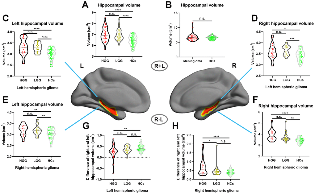
Figure 2.Increased hippocampal volume in glioma patients with ROI-based analysis. (A) Hippocampal volume significantly increased in both the HGG and LGG groups compared to HCs (one-way ANOVA with Bonferroni correction, **** p < 0.0001, n.s. represents no significance). (B) No significant difference in hippocampal volume between meningioma patients and HCs (two-sample t test, p = 0.24). (C, D) Right and left hippocampal volume significantly increased in left hemispheric glioma (including left LGG and left HGG) compared to HCs (one-way ANOVA with Bonferroni correction, * p < 0.05, *** p < 0.001, **** p < 0.0001). (E, F) Right and left hippocampal volume significantly increased in right hemispheric glioma (including right LGG and right HGG) compared to HCs (one-way ANOVA with Bonferroni correction, ** p < 0.01, **** p < 0.0001). (G, H) For right and left hemispheric high-grade glioma patients, there was a significant increase in the ipsilateral hippocampal volume relative to the contralesional hippocampal volume in the HGG group compared to the LGG and HCs groups (one-way ANOVA with Bonferroni correction, ** p < 0.01, **** p < 0.0001). L: left hemisphere, R: left hemisphere.