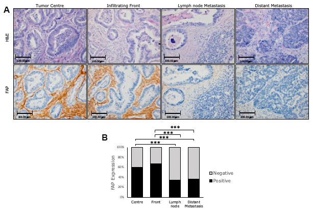
Figure 3.Immunohistochemical FAP expression in primary (centre and border) and corresponding metastatic (lymph node and liver) tissues of conventional adenocarcinomas (AdCs). (A) Higher percentage of positive staining was observed in primary tumours than in metastases (x200). (B) FAP staining intensity was scored as negative or positive. The scores were quantified in each tissue type and statistical significance of FAP expression pattern among the different tissues was determined by Chi-Square test (***p<0.001). H&E: Hematoxylin and Eosin staining. FAP: Fibroblast activation protein-α.