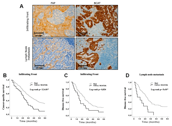
Figure 4.Immunohistochemical FAP and nuclear BCAT staining in the infiltrating front and in lymph node metastasis. (A) FAP was expressed in CAFs that penetrated within the body of lymphatic nodes. Nuclear and adjacent cytoplasmic BCAT staining represents BCAT signalling translocation from membrane to nucleus (x200). (B, C) Kaplan-Meier curves and univariate Log-rank test showed that simultaneous expression of FAP in CAFs and nuclear BCAT in AdC cells from the infiltrating front significantly associated with worse 5-year cancer-specific (CSS) and disease-free (DFS) survival of AdC patients. (D) The same combination in lymph node metastases was significantly associated to 5-year DFS.