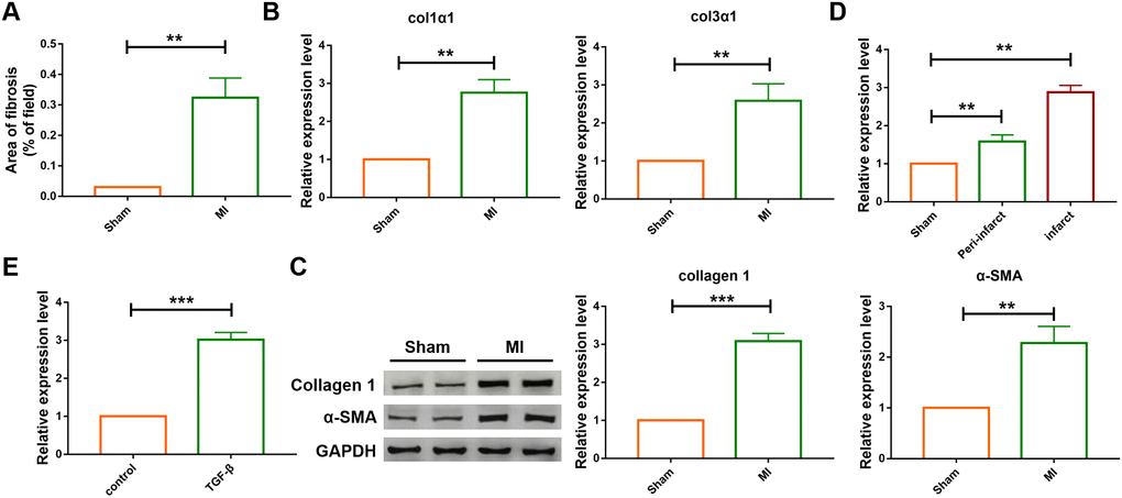
Figure 1.The differential expression of SNHG7 in cardiac tissues and cardiac fibroblast. (A) Quantification of the total fibrotic area using Image-J. Fibrosis areas of sham-operated group and MI group were detected by Masson staining. Data was presented as mean ± SEM; two-tailed t test was used for the statistical analysis. n=7 mice per group. (B) mRNA expression of collagen 1α1 and collagen 3α1 were measured by qRT-PCR; GAPDH mRNA served as an internal control. Data was presented as mean ± SEM; two-tailed t test was used for the statistical analysis. n=6 mice per group. (C) Protein levels of collagen 1 and α-SMA were measured by western blotting analysis; GAPDH served as an internal control. Data was presented as mean ± SEM; two-tailed t test was used for the statistical analysis. n=6 mice per group. (D) qRT-PCR analysis showing upregulation of lncRNA SNHG7 in the peri-infarcted and infarcted areas of left ventricle of mice after MI. Data was presented mean ± SEM; two-tailed t test was used for the statistical analysis. n=5 mice per group. (E) qRT-PCR analysis showing elevation of lncRNA SNHG7 in cardiac fibroblasts after treatment with TGF-β1 (10 ng/mL) for 24h. Data was presented as mean ± SEM; two-tailed t test was used for the statistical analysis. n=5 independent cell cultures. **P<0.05, ***P<0.001.