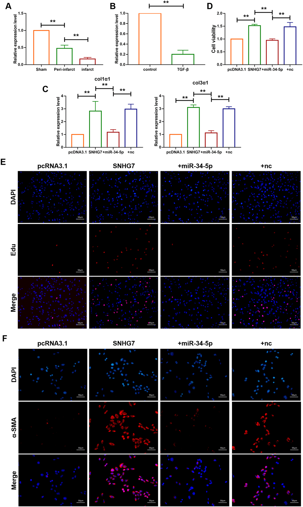
Figure 4.lncRNA SNHG7 promoted cardiac fibrosis by targeting miR-34-5p. (A) qRT-PCR analysis showing downregulation of miR-34-5p in the peri-infarcted and infarcted areas of left ventricle of mice after MI. U6 served as an internal control. Data was presented as mean ± SEM; two-tailed t test was used for the statistical analysis. n=5 mice per group. (B) qRT-PCR analysis showing reduction of miR-34-5p in cardiac fibroblasts after treatment with TGF-β1 (10 ng/mL) for 24h. Data was presented as mean ± SEM; two-tailed t test was used for the statistical analysis. n=5 independent cell cultures. (C) mRNA expression of collagen 1α1 and collagen 3α1 were measured by qRT-PCR. Forced expression of SNHG7 (1 μg/mL) in cardiac fibroblasts increased mRNA expression levels of collagen 1α1 and collagen 3α1, which were reversed by miR-34-5p overexpression; GAPDH mRNA served as an internal control. Data was presented as mean ± SEM; one-way ANOVA was used for the statistical analysis. n=6 mice per group. (D) MTT assay for the assessment of cell viability. Transfection of SNHG7 with or without miR-34-5p in normal cardiac fibroblasts. Data was presented as mean ± SEM; two-tailed t test was used for the statistical analysis. n=5 independent cell cultures. (E) EdU staining for the assessment of cell proliferation in cardiac fibroblasts overexpressing SNHG7 in the presence or absence of miR-34-5p mimics. Scale bars represented 50 μm. (F) Representative images of immunofluorescence staining showing that forced expression of SNHG7-induced fibroblast-myofibroblast transition. Scale bars represent 50 μm. **P<0.05.