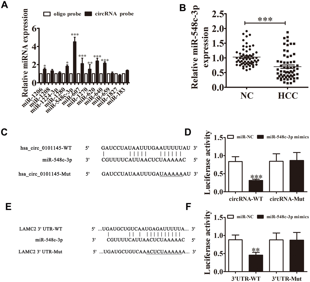
Figure 5.Interactions among hsa_circ_0101145, miR-548c-3p, and LAMC2. (A) RT-qPCR analyses revealed that miR-548c-3p was the only miRNA that was abundantly pulled down by the hsa_circ_0101145 probe in HepG2 cells. Data are presented as means ± SD. *P < 0.05, **P < 0.01, ***P < 0.001 vs oligo. (B) RT-qPCR shows the expression of miR-548c-3p in HCC tissues (60) and adjacent normal tissues (60). Data are presented as the mean ± SD. ***P < 0.001 vs. Normal. (C) Prediction of binding sites of miR-548c-3p in hsa_circ_0101145. The mutant version of hsa_circ_0101145 is presented. (D) Relative luciferase activity determined at 48 h after transfection of 293T cells with miR-548c-3p mimic/NC or hsa_circ_0101145 wild-type/Mut. Data are presented as means ± SD. ***P < 0.001. (E) Prediction of binding sites of miR-548c-3p in the 3'UTR of LAMC2. The mutant version of 3'UTR-LAMC2 is shown. (F) Relative luciferase activity determined at 48 h after transfection of 293T cells with miR-548c-3p mimic/NC or 3'UTR-LAMC2 wild-type/Mut. Data are presented as means ± SD. ***P < 0.001.