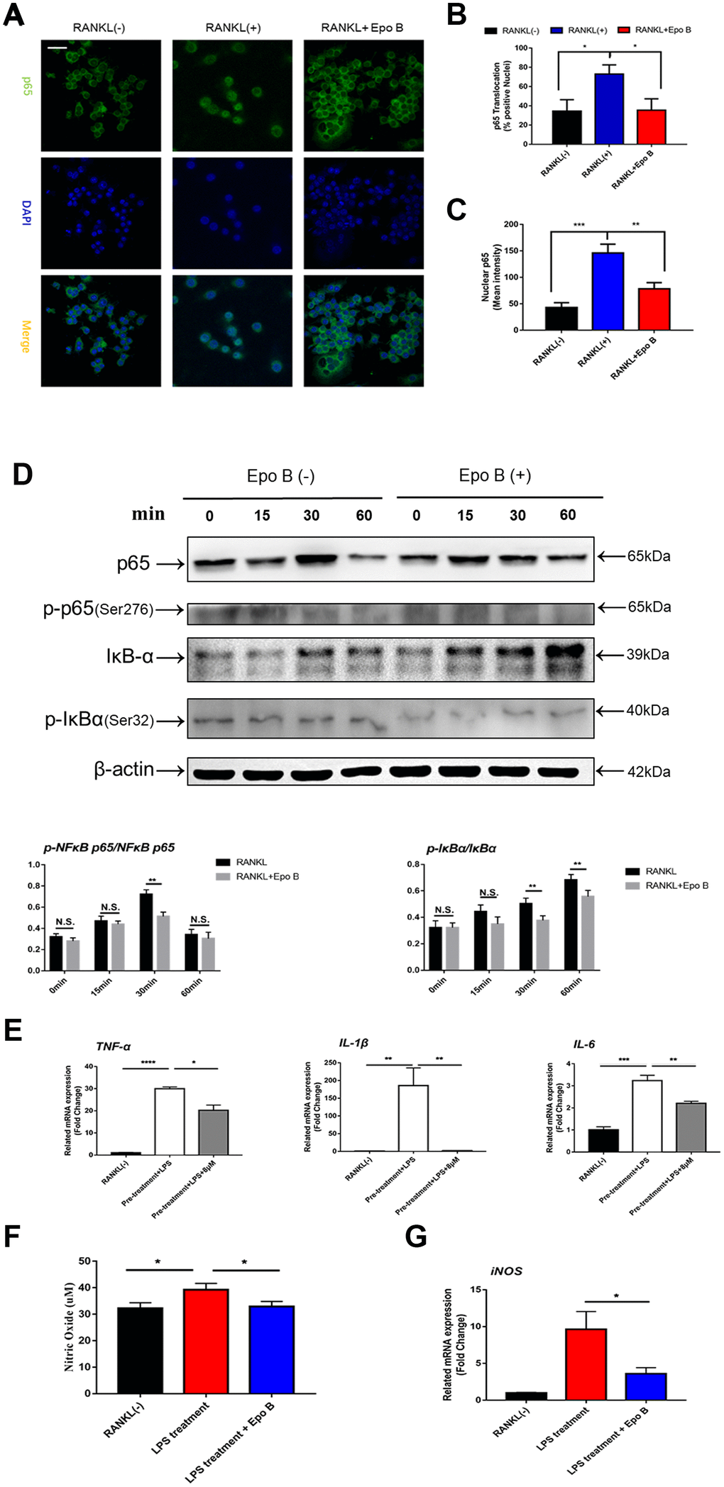
Figure 5.Epothilone B restrained NF-ĸB signaling pathway and inhibited the release of pro-inflammatory cytokines and nitric oxide. (A) Representative images of immunofluorescence staining of the nuclear translocation of NF-ĸB p65 in the absence of presence of Epothilone B. Scale bar = 400 μm. (B) Quantitative analysis of the percentage of positive cells (NF-ĸB p65 translocation from cytosol to nuclear) in all cells. (C) Quantitative analysis of the mean intensity of NF-ĸB p65 in the cells nuclear. (D) RAW264.7 cells were stimulated with RANKL with or without Epothilone B (8μM) for the 0-60 minutes. The cell lysates were analyzed using western blotting for p-NFκB p65, NFκB p65, p-IκBα and IκBα. Quantification protein expression of p-NFκB p65 relative to NFκB p65 and p-IκBα relative to IκBα. (E) Relative expression of pro-inflammatory cytokines (TNF-α, IL-1β, IL-6) during LPS induced osteoclast differentiation on mRNA level. (F) The concentration of NO was detected in the process of LPS-induced osteoclastogenesis. (G) Relative expression of iNOS (inducible nitric oxide synthetase) during LPS induced osteoclastogenesis on mRNA level. Data in the figures represent mean ± SD. N.S. represented no significant difference. *p < 0.05, **p < 0.01, ***p < 0.001 based on one way ANOVA.