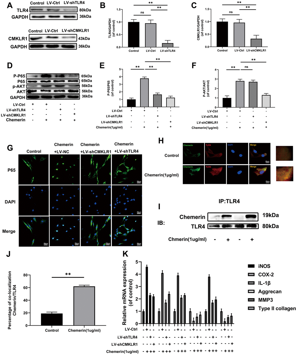
Figure 5.TLR4, and CMKLR1 were involved in chemerin-induced signaling pathway activation, ECM disorder and inflammatory response. (A) The expression levels of TLR4, and CMKLR1 were visualized by western blotting. (B, C) Quantification of TLR4, and CMKLR1 immunoblots. (D) The expression levels of p-AKT, AKT, p-p65, and p65 were evaluated by western blotting. (E, F) Quantification of p-AKT, AKT, p-p65, and p65 immunoblots. (G) The expression levels of p65, and nuclear translocation were observed by immunofluorescence. (H) Representative image of immunofluorescence double staining of TLR4, and chemerin in NPCs. (J) The quantification of the percentage of co-location of chemerin/TLR4 was detected by image J. (I) The co-immunoprecipitation data showed that compared with untreated group, the binding of chemerin to TLR4 was significantly increased after treatment with chemerin. (K) The mRNA expression levels of iNOS, COX-2, IL-1β, aggrecan, MMP3, and collagen II in NPCs were evaluated by RT- PCR. Data are represented as mean ± SEM of three independent experiments, each done in triplicate. Significant differences between groups are indicated as **p < 0.01, *p < 0.05.