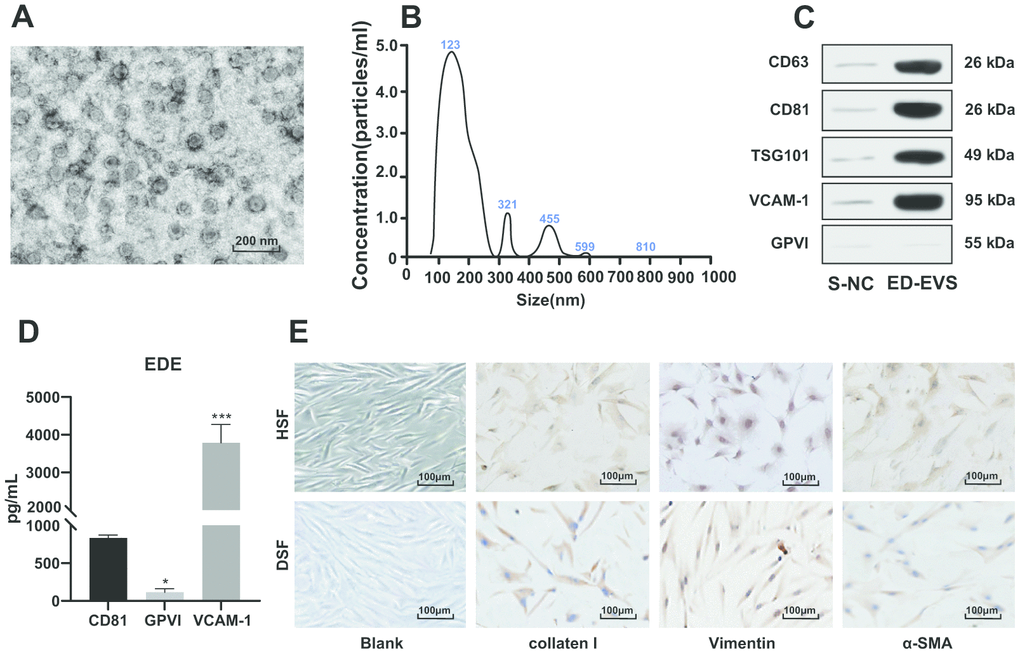
Figure 1.Identification of plasma ED-EVs and skin fibroblasts. (A) The morphology and size of EVs was observed by transmission electron microscope. (B) The nanoparticle tracking software showed that the average diameter of ED-EVs was 123 ± 8 nm, and the concentration was 5.2 × 10⁷ particles/mL. (C) Western blot analysis showed that CD63, CD81, TSG101 and VCAM-1 were higher in plasma ED-EVs than those in S-NC, and GPVI was markedly low in both groups. S-NC is the supernatant after immunoprecipitation. (D) The amount of proteins carried in ED-EVs was quantified by ELISA; compared with CD81, * p < 0.05, *** p < 0.001. (E) Collagen I and Vimentin were positive and α-SMA was weakly positive as immunocytochemistry indicated. Data in panel C were analyzed by two-way ANOVA, and in panel D were analyzed by one-way ANOVA, followed by Tukey's multiple comparisons test. Repetitions = 3.