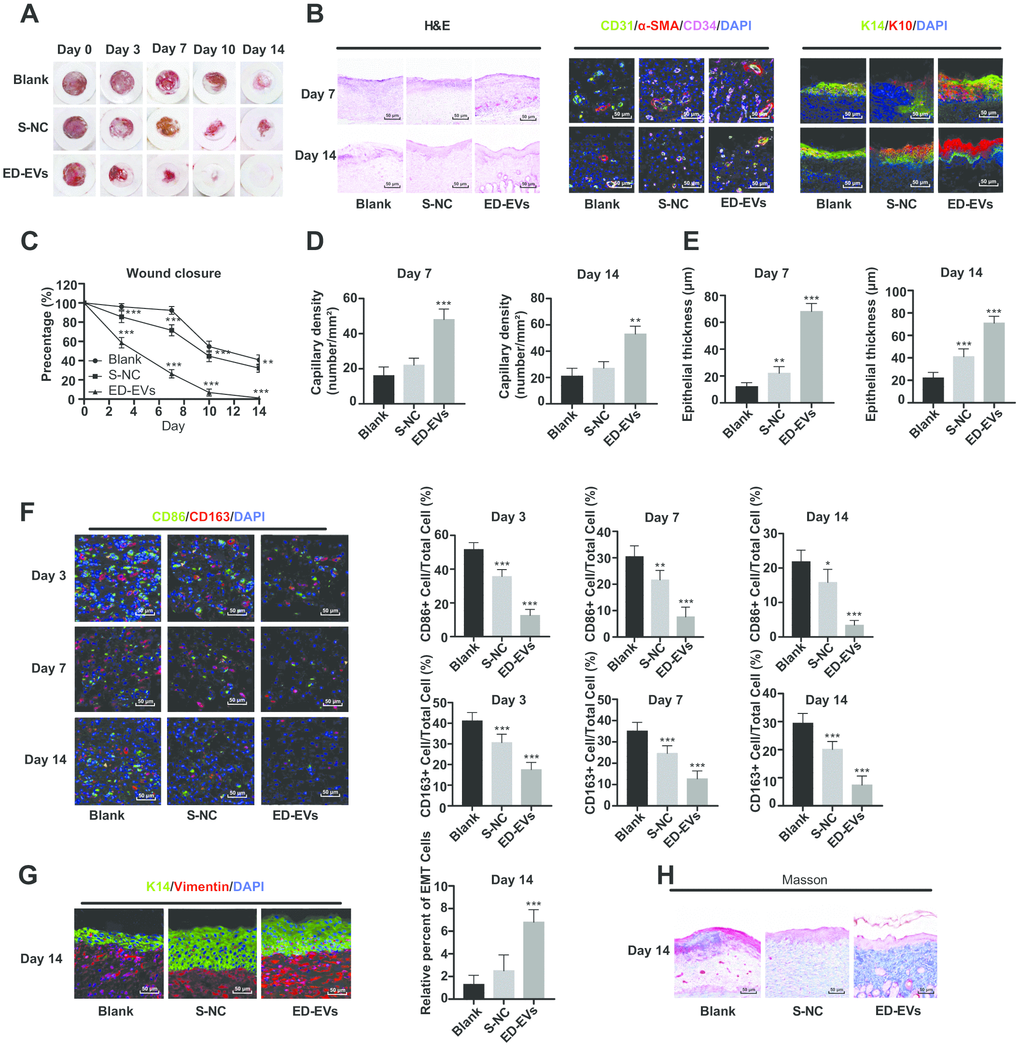
Figure 6.Plasma ED-EVs promote skin wound healing in diabetic mice. (A and C) The wound area of diabetic mice was measured on the 0, 3, 7, 10 and 14 days after skin wound modeling (n = 6). On the 3rd, 7th and 14th day after skin wound modeling, the pathological changes (B, H), vascular markers (B, D), re-epithelialization (E), macrophage infiltration and EMT (F, G) and collagen deposition (F) in the skin of diabetic mice were detected by HE staining (n = 3), immunofluorescence (n = 6) and Masson staining (n = 3). Compared with the blank group, *p < 0.05, **p < 0.01, ***p < 0.001. Data in panel (C) were analyzed by two-way ANOVA, and data in panels (D–G) were analyzed by one-way ANOVA, followed by Tukey's multiple comparisons test. Repetitions = 3.