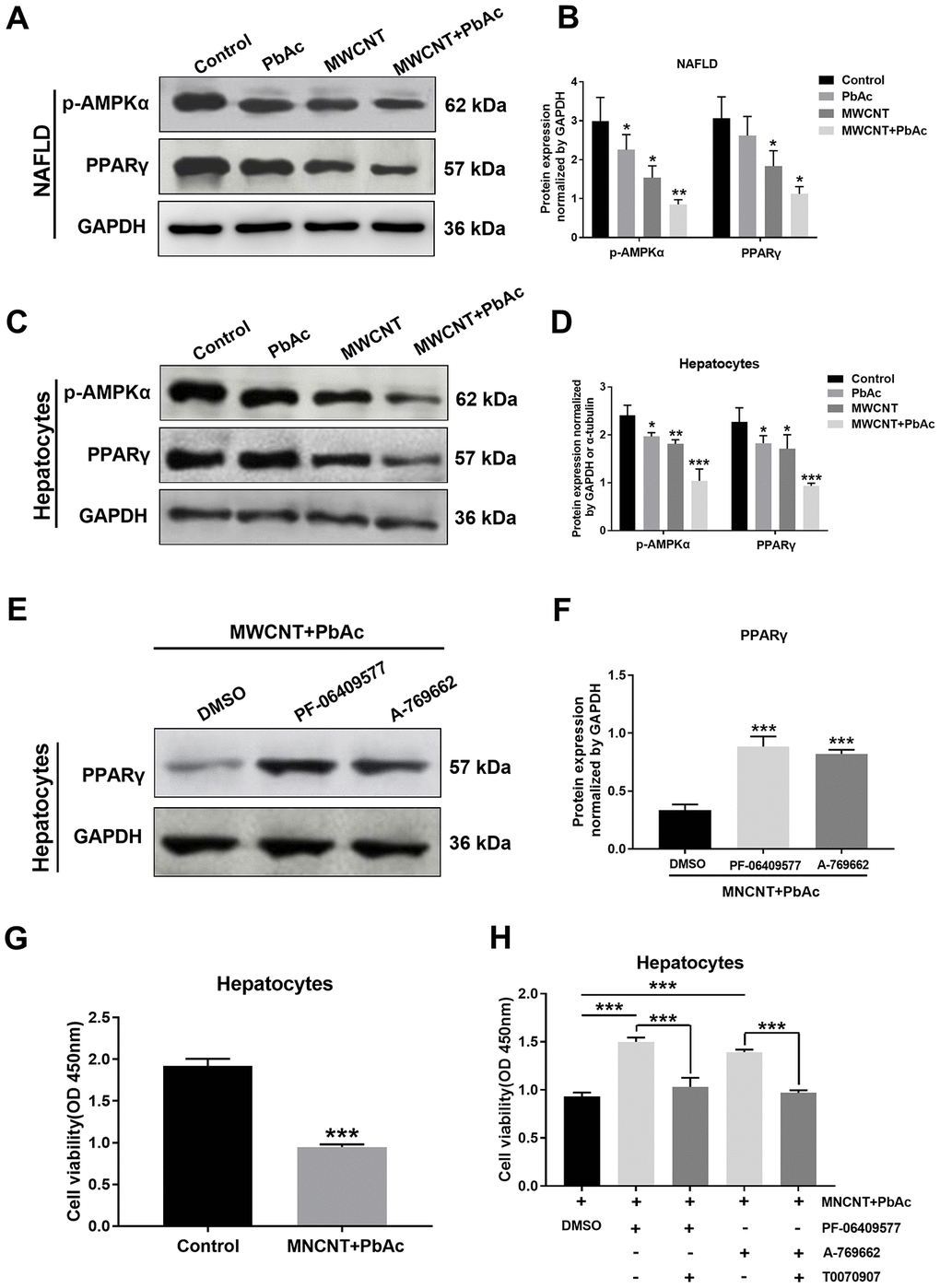
Figure 9.Combined administration of MWCNTs and PbAc may exert its hepatotoxicity to NAFLD mice via inhibiting AMPK/PPARγ pathway. Western blot analysis of PPARγ and p-AMPKα expressions in NAFLD mice livers (A) and in primary hepatocytes from NAFLD mice (C) upon the low dose of PbAc, MWCNTs or MWCNTs + PbAc administration. (B, D) PPARγ and p-AMPKα expression levels normalized to GAPDH (*P<0.05, **P<0.01 and ***P<0.001, compared to saline water). (E) After treatments with DMSO or two AMPK activators (0.5 μM PF-06409577 and 10 μM A-769662) in addition to MWCNTs + PbAc, PPARγ expressions in primary hepatocytes from NAFLD mice were tested using western blot analysis. (F) PPARγ expression levels normalized to GAPDH (***P<0.001, compared to DMSO). (G) Cell viability of primary hepatocytes from NAFLD mice upon the administration of saline water or MWCNTs + PbAc (***P<0.001, compared to saline water). (H) In addition to MWCNTs + PbAc, primary hepatocytes from NAFLD mice were also incubated with DMSO, PF-06409577 (0.5 μM), A-769662 (10 μM) or T0070907 (a selective PPARγ antagonist, 50 μM), then the cell viability was measured (***P<0.001, compared to DMSO or T0070907).