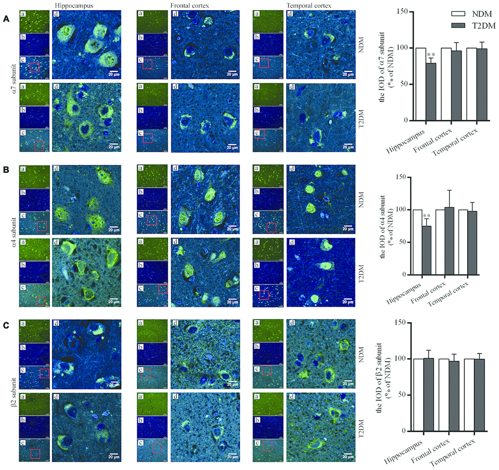
Figure 1.Immunofluorescent staining for nAChR α7 (A), α4 (B) and β2 (C) subunits in the hippocampus (CA3), and the frontal and temporal cortices of patients with T2DM (n=6) and age-matched controls (NDM, n=6). Photographs were taken by using a laser confocal microscope. The α7, α4 and β2-positive neurons were reacted by specific antibodies as shown as green (a); cell nuclei are stained as blue (using DAPI) (b); a and b were merged as one picture (c); and a partial area from c was selected to be magnified (d) with scale bars=20 μm. The values presented as percentage of the control by relative quantification for α7, α4 or β2 subunit staining in those regions are means ± SEM; **p<0.01 as compared to NDM employing the two-tailed unpaired Student’s t test.