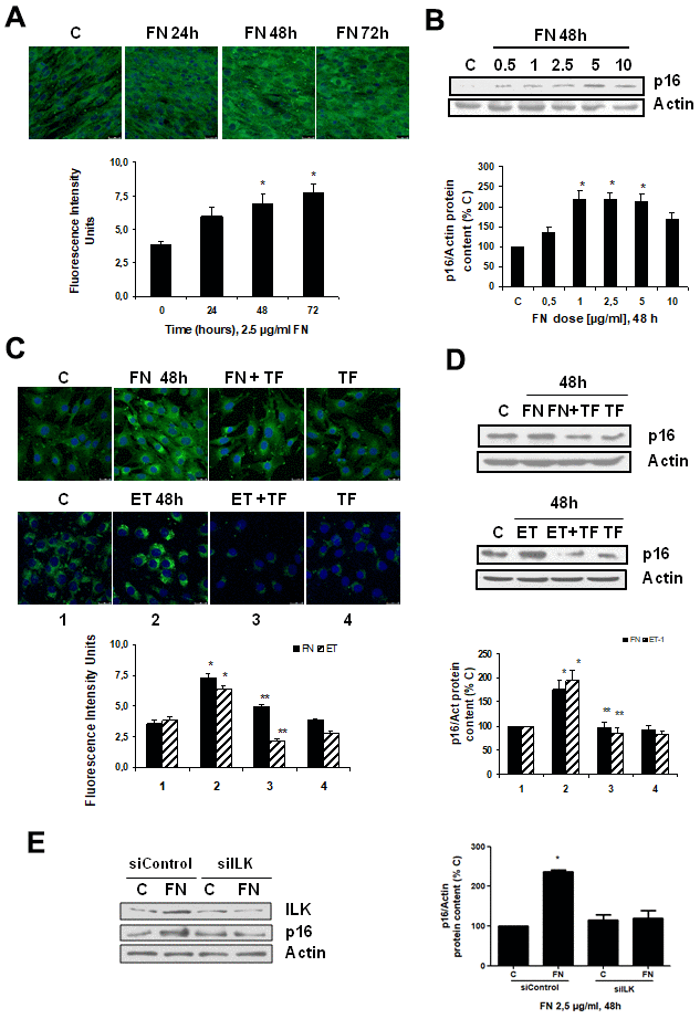
Figure 3.Fibronectin induces senescence in mouse myoblasts (C2C12) through integrin/ILK activation. Cells were grown on coverslips (panels A, C) to test senescence measuring SA-ß-GAL activity and p16 protein content by Western blot (panels B, D). (A) Cells were incubated with 2.5 μg/ml FN at different times to assess SA-ß-GAL activity by confocal microscopy. (B) Cells were incubated at different doses of FN for 48h to analyze p16 protein content. (C) Cells were incubated with 2.5 μg/ml FN or 1 nM ET-1 in the presence or not of 50 μM Tirofiban (TF) for 48h to assess senescence by SA-ß-GAL activity (panel C) or by p16 protein content (panel D). Representative microphotographs are shown at the top with 40x magnification and the densitometric analysis is shown below. Scale bar, 50 μm. A representative Western blot of p16 is shown at the top and the densitometric analysis is shown below. In panels C and D closed bars represent data of FN treatment and stripped bars represent data of ET-1 treatment; lane 1: control cells; lane 2: FN or ET alone; lane 3: FN or ET plus TF; lane 4: TF alone. Values are the mean±SEM of 6 independent experiments, *p<0.05 vs. control cells (C or time 0), and **p<0.05 vs ET or FN alone. (E) Cells were transfected with siRNA against ILK or scrambled as siControl to assess senescence by p16 protein content upon 2.5 μg/mL FN treatment for 48h. A representative Western blot of ILK and p16 are shown on the left panel and the densitometric analysis is shown on the right. Values are the mean±SEM of 3 independent experiments, *p<0.05 vs. control cells (C from siControl).