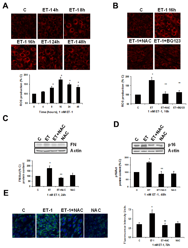
Figure 4.Role of ROS in endothelin-dependent fibrosis and cellular senescence. (A, B) Cells were incubated with 1 nM ET-1 at different times, some cells were incubated in the presence of 100 μM N-acetylcysteine (NAC) or 100 nM BQ123, and then 1 nM ET-1 was added and incubated for 16h (B). CellROX probe was added during the last 30 min of incubation. After being washed twice, in vivo cells were visualized by microscopy confocal to test ROS production in red. Representative microphotographs are shown at the top with 40x magnification, scale bar, 50 μm. The densitometric analyses are shown below. (C–E) Cells were incubated with 1 nM ET-1 in the presence of 100 μM NAC to assay FN protein expression by Western blot (C). Cellular senescence was assessed by measuring p16 protein content for 48h by Western blot (panel D) and SA-ß-GAL activity for 72h (panel E). A representative Western is shown on the top with the densitometric analysis below (panels C, D). Representative microphotographs are shown on the left panel E with 40x magnification and the densitometric analysis is shown on the right. Scale bar, 50 μm. Values are the mean±SEM of 6 independent experiments, *p<0.05 vs. control cells (C or time 0), and **p<0.05 vs ET alone.