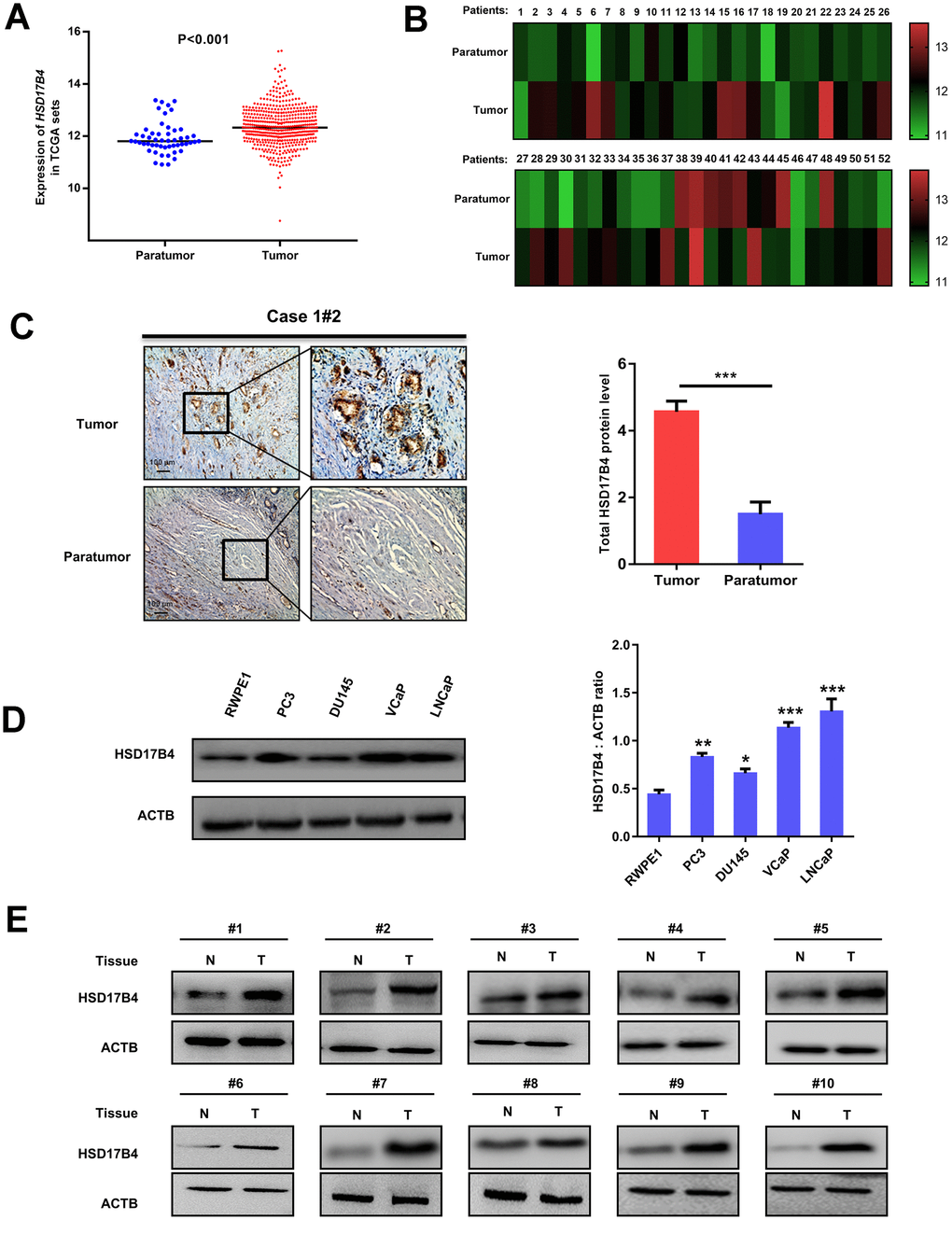
Figure 1.Expression of HSD17B4 is increased as PCa develops. (A) The expression of HSD17B4 was frequently upregulated in 498 PCa tissues (Tumor) compared with 52 adjacent normal prostate tissue samples (Paratumor) in the TCGA profile. (B) HSD17B4 expression was markedly increased in 52 paired PCa tissues (Tumor) and their adjacent normal tissues (Paratumor) in the TCGA profile. (C) Immunohistochemical staining of total HSD17B4 protein in tumor and adjacent tissues. A total of 10 PCa tissues and 10 adjacent normal prostate tissues were analyzed. Quantitative analysis of total HSD17B4 expression was performed by ImageJ. (D) Characterization of total HSD17B4 protein levels in PCa cell lines. Equal amounts of protein (20 μg) were immunoblotted for HSD17B4 and ACTB (loading control), as shown in the left panel. The intensities of total HSD17B4 protein are quantified in the right panel. (E) HSD17B4 is overexpressed in PCa tissues compared to expression in adjacent tissues. Human PCa samples each paired with cancerous tissue (designated as T) and adjacent normal tissue (designated as N) were lysed and directly subjected to western blotting. Ten pairs of samples clearly exhibited HSD17B4 overexpression in PCa tissues. Data are shown as the mean ± SD (n = 3) or typical photographs of one representative experiment. Similar results were obtained in three independent experiments. *p < 0.05, **p < 0.01, ***p <0.001.