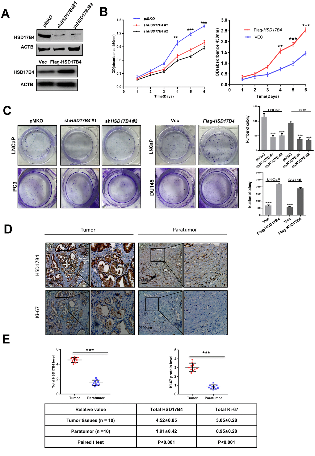
Figure 2.HSD17B4 promotes the proliferation of PCa cells. (A) Verification of LNCaP stable cell lines. The transfection efficiencies of shHSD17B4 and Flag-HSD17B4 were determined by western blotting. (B) HSD17B4 knockdown inhibited LNCaP cell growth, while HSD17B4 overexpression promoted cell growth. The CCK-8 assay showed that the growth of LNCaP cells characterized in (A) was affected by HSD17B4 knockdown or overexpression. The data shown are representative of three independent experiments. (C) HSD17B4 promotes the proliferation of PCa cells. PCa cells were transfected with shHSD17B4 or Flag-HSD17B4 plasmids as indicated and analyzed by a colony formation assay (left panel). Quantitative analysis of the colony was performed by ImageJ. **denotes P < 0.01; ***denotes P< 0.001. Error bars represent ±SD of triplicate experiments (right panel). (D–E) Immunohistochemical staining of Ki-67 and total HSD17B4 protein in tumor and adjacent tissues. Examples are shown in (D), and the statistical analysis of all samples is shown in (E). Scale bars: 100 μm. The intensities of the HSD17B4 and Ki-67 proteins in 10 PCa tissues (upper panel) and 10 adjacent normal prostate tissues (lower panel) were quantified, followed by statistical analysis. The mean value of multiple samples and the standard deviation are presented. Data are shown as the mean ± SD (n = 3) or typical photographs of one representative experiment. Similar results were obtained in three independent experiments. *p < 0.05, **p < 0.01, ***p <0.001.