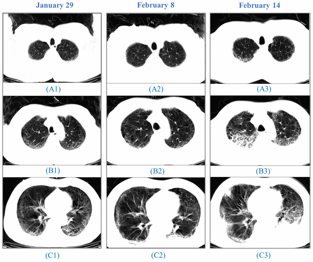
Figure 2.CT images from a 64-year-old man. A 64-year-old man, who had a fever and pneumonia, was suspected as the SARS-CoV-2 carrier on January 28 and confirmed on January 30. (A1), (B1) to (C1): On January 29, initial CT scans at the hospital admission showed multifocal ground-glass opacity (GGO) and reticulation, predominantly in the subpleural areas of both lungs. (A2), (B2) to (C2): On February 8, CT images indicated progressing GGOs. Newly-appeared patchy and core-like consolidation were visible in lower lobes of both lungs. The patient showed high fever, cough, blood in the sputum, reduced SpO2, and a sign of heart failure. (A3), (B3) to (C3): On February 14, CT images showed progressing lesion with multiple newly-appeared GGO and consolidation. Irregular interlobular septal thickening was observed in the upper lobe of the right lung. The patient passed away on February 15.