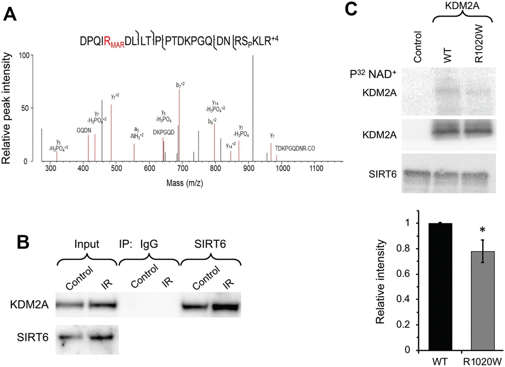
Figure 1.SIRT6 mono-ADP ribosylates KDM2A on R1019/R1020 (mouse/human). (A) Mass spec analysis of mouse KDM2A mono-ADP ribosylation. MS2 fragment spectrum of a KDM2A peptide obtained from mouse embryonic fibroblast cells supporting R1019 modified by a ribose-phosphate group indicating mono-ADP ribosylation (MAR). Pictured is an assignment showing ribose-phosphate modified R1019. A full compiled report downloaded from Protein Prospector of the MS2 data for this peptide can be found in the Supplementary Data (Supplementary Data 1). (B) SIRT6 interacts with KDM2A. coimmunoprecipitation of KDM2A was observed with antibodies directed against SIRT6 before and after irradiation (IR) (the experiment was repeated three times). (C) SIRT6 mono-ADP ribosylates KDM2A in vitro. KDM2A R1020W mutation significantly decreases SIRT6 mono-ADP ribosylation signal. Top panel. SIRT6 protein was incubated with purified human wild type flag-KDM2A or mutant R1020W proteins in the presence of radiolabeled NAD+. Mono-ADP ribosylation was detected by transfer of the radiolabel to the substrate. Lower panel. Quantification of upper gel. Graph represents mean ± SD of three experiments. *p > 0.05.